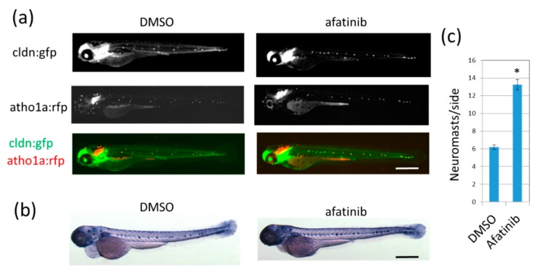
Afatinib induces the development of extra lateral line neuromasts in zebrafish larvae. (a,b) Neuromasts of 72 hpf larvae treated with DMSO (left) or afatinib (right) from 26 hpf were detected by fluorescence from cldn:gfp; atoh1:rfp transgenic larvae (a) or by endogenous ALP activity (b). (a) Fluorescence from cldnb:gfp highlighted neuromast cells and the epidermis, and fluorescence from atoh1a:rfp highlighted neuromast hair cells. GFP and RFP images were merged. Compared to DMSO treatment, increased neuromasts were observed in afatinib-treated zebrafish larvae. Scale bar = 500 µm. (c) The number of neuromasts on one side were counted. The graph indicates mean ± SEM (n = at least 54). * p < 0.01; hpf, hours postfertilization; DMSO, dimethyl sulfoxide; ALP, alkaline phosphatase; GFP, green fluorescent protein; RFP, red fluorescent protein; SEM, standard error of the mean.
Acknowledgments
This image is the copyrighted work of the attributed author or publisher, and
ZFIN has permission only to display this image to its users.
Additional permissions should be obtained from the applicable author or publisher of the image.
Full text @ Pharmaceuticals (Basel)
Your Input Welcome
Thank you for submitting comments. Your input has been emailed to ZFIN curators who may contact you if
additional information is required.
Oops. Something went wrong. Please try again later.