
- Title
-
Human macrophages survive and adopt activated genotypes in living zebrafish
- Authors
- Paul, C.D., Devine, A., Bishop, K., Xu, Q., Wulftange, W.J., Burr, H., Daly, K.M., Lewis, C., Green, D.S., Staunton, J.R., Choksi, S., Liu, Z.G., Sood, R., Tanner, K.
- Source
- Full text @ Sci. Rep.

Human macrophages survive in vivo for up to two weeks post-injection following brain injection. (a) Schematic of experimental design: primary monocytes were differentiated into macrophages before injection into the zebrafish brain at age 2 days post fertilization (dpf) and imaged at 1, 7 and 14 days post injection (dpi). (b) Micrographs of representative whole larva at 3 dpf (left) and 3D projections showing distribution and survival of human primary macrophages (blue) injected into the hind brain of transgenic mpx:GFP (neutrophils-green)/flk:mCherry (vessels-red) zebrafish larvae at 1 dpi (3 dpf) (right). (c) Micrographs of representative whole larva at 9 dpf (left) and 3D projections showing distribution and survival of human primary macrophages (blue) injected into the hind brain of transgenic mpx:GFP (neutrophils-green)/flk:mCherry (vessels-red) zebrafish larvae at 7 dpi (9 dpf) (right). (d) Micrographs show that cells can persist for up to 2 weeks after injection at 16 dpf. Top panel: representative zebrafish at 16 dpf. Left panel: micrograph shows tiled image of transgenic mpx:GFP (neutrophils-green)/flk:mCherry (vessels-red) 16 dpf zebrafish, white square highlights region of interest in the zebrafish brain. Right panel: micrograph of the inset where the white arrows indicate human cells. Scales are indicated on each image.
|

Human macrophages survive and interact with zebrafish stromal cells post-injection following brain injection. (a) Schematic of experimental design: primary monocytes were differentiated into macrophages before injection into the zebrafish brain at age 2 days post fertilization (dpf) and imaged at 1 and 2 days post injection (dpi). (b) 3D projections of whole head at 3 dpf (left) and 4 dpf (middle) showing distribution and survival of human primary macrophages (blue) injected into the hind brain of flk:mCherry (vessels-red) (top) and GFAP:GFP (astrocytes-green)/flk:mCherry (vessels-red) (bottom) zebrafish larvae. Boxes indicate positions of insets shown in right panels. Scales are indicated.
|

Human macrophages survive in vivo for up to two weeks post-injection following injection directly into the circulation of the zebrafish. (a) Schematic showing experimental set up, where human macrophages were directly injected into the circulation of 2 days post-fertilization (dpf) larvae, after being differentiated from primary human monocytes. Injected larvae were imaged at 1, 7 and/or 14 days post injection (dpi). (b,c) Micrographs of whole larva at 3 dpf (left) showing distribution and survival of human primary macrophages (blue) differentiated at either at physiological temperature of zebrafish (28.5 °C) or at physiological temperature of humans (37 °C) before injection into the caudal vein of transgenic fli:GFP (vessels-red) zebrafish larvae. Micrographs display macrophage distribution at 1 dpi. Bottom panels: Micrographs of 3D projections at higher magnification of insets highlighted in tiled images showing distribution and survival of human primary macrophages (blue). (d) Top panel: micrograph shows tiled image of the 9 dpf zebrafish, 7dpi, showing distribution and survival of human primary macrophages differentiated at human physiological temperature (37 °C) (blue) injected into transgenic fli:GFP (vessels-red) zebrafish larva. Bottom panel: Micrographs of 3D projections at higher magnification of insets highlighted in tiled image showing distribution and survival of human primary macrophages (blue). (e) Top panel: micrograph shows representative tiled image of a 16 dpf zebrafish. Bottom panel: Micrographs of 3D projections showing distribution and survival of human primary macrophages (blue) at 14 dpi following injection to transgenic mpx:gfp (neutrophils-green)/flk:mCherry (vessels-red) zebrafish larvae at 2 dpf. Arrows indicate macrophage positions.
|

Primary human monocytes survive at physiological temperature of the zebrafish in vitro and in vivo. (a) Primary human monocytes (blue) were injected into the hind brain of transgenic GFAP:GFP (astrocytes-green)/flk:mCherry (vessels-red) zebrafish larvae at 2 dpf. Micrographs show representative images where zebrafish was imaged serially over the course of 3 days post-injection at interval of 24 hours. (b) Plot (mean ± 95% CI) of average in vivo survival calculated for primary human monocytes obtained from larvae where the numbers of cells that survived over the course of 3 days for each larvae were normalized to the initial numbers measured one day post injection. Statistical analysis where * indicates a p value of p < 0.025 and *** indicates a p value of p < 0.0005 by successive t tests with Bonferroni correction, with n = 41 larvae for Day 1, n = 41 larvae for Day 2, and n = 4 larvae for Day 3. Scatter plots show values for individual zebrafish. Not all zebrafish survived to 5 days post injection. (c) Cells were either cultured at physiological temperature of humans (37 °C) or at physiological temperature of zebrafish (28.5 °C). Plot (mean ± SD) of average survival calculated for primary human monocytes obtained from n = 4 donors where the numbers of cells that survived over the course of 7 days were normalized to the original seeding density. Days in culture was a statistically significant factor in survival by two-way ANOVA with one degree of freedom (P < 0.0001), as was the temperature of culture (P = 0.0018).
|
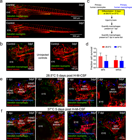
Primary human macrophages can survive at physiological temperature of the zebrafish in vivo and do not cause a sustained inflammatory response. (a) Representative light sheet micrographs of uninjected, control whole larva at 3 days post-fertilization (dpf). Both transgenic mpeg:GFP (macrophages-green)/flk:mCherry (vessels-red) (top) and mpx:GFP (neutrophils-green)/flk:mCherry (vessels-red) (bottom) are shown. (b) Representative lightsheet micrographs of head regions of uninjected, control larvae at 3 dpf. Both transgenic mpeg:GFP (macrophages-green)/flk:mCherry (vessels-red) (top) and mpx:GFP (neutrophils-green)/flk:mCherry (vessels-red) (bottom) are shown. (c) Schematic of experimental design: primary monocytes were differentiated into macrophages before injection into the zebrafish brain at age 2 dpf and imaged at 1 and 7 days post injection (dpi). (d) Plot (mean ± SD) of average in vivo survival calculated for primary human macrophages differentiated at physiological temperatures of the zebrafish or human obtained from 3 larvae each, where the numbers of cells that survived over the course of 7 days were normalized to the initial numbers measured one day post injection. Differences in survival were not significant by two-way ANOVA with Tukey’s multiple comparisons post-test. The temperature of macrophage differentiation (P = 0.3072), fish line injected (P = 0.4822), and interaction between factors (P = 0.4575) were not significant sources of variation by two-way ANOVA with one degree of freedom. Monocytes that had been cultured in the presence of human macrophage colony stimulating factor (H-M-CSF) for eight days (e) at physiological temperature of (28.5 °C) and (f) at physiological temperature of humans (37 °C). Primary human macrophages (blue) were injected into the hind brain of transgenic mpeg:GFP (macrophages-green)/flk:mCherry (vessels-red) or mpx:GFP (neutrophils-green)/flk:mCherry (vessels-red) zebrafish larvae at 2 dpf. Micrographs of 3D projections showing distribution and survival of human primary macrophages 1 dpi, when larvae are 3 dpf, and 7 dpi, when larvae are 9 dpf. The same fish is shown in the left and right panels.
|