
- Title
-
Mutation of microphthalmia-associated transcription factor (mitf) in zebrafish sensitizes for glomerulopathy
- Authors
- Müller-Deile, J., Schenk, H., Niggemann, P., Bolaños-Palmieri, P., Teng, B., Higgs, A., Staggs, L., Haller, H., Schroder, P., Schiffer, M.
- Source
- Full text @ Biol. Open
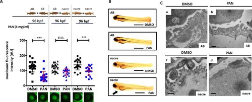
Influence of genetic background on the proteinuria phenotype. (A) Tg(l-fabp:eGFP-DBP) zebrafish backcrossed on homozygous for either the AB (AB fish) or the nacre (nacw2) background (nacre fish) as well as heterozygous zebrafish for nacre and AB background were treated with PAN at 46 h post-fertilization (hpf). Graph depicts max. fluorescence of circulating eGFP-DBP in the retinal vessel plexus of the fish eye 96 hpf. (B) Representative images of phenotypes of Tg(l-fabp:eGFP-DBP) zebrafish backcrossed on homozygous for either the AB (AB fish) or the nacre (nacw2) background (nacre fish) were treated with PAN (4 mg/ml) or DMSO at 46 hpf as indicated. Pictures were taken at 96 hpf. (C) Representative transmission electron microscopy images of the glomerular filtration barrier of Tg(l-fabp:eGFP-DBP) zebrafish backcrossed on homozygous for either the AB or the nacre (nacw2) background were treated with PAN (4 mg/ml) or DMSO at 46 hpf as indicated. Fish were collected at 120 hpf. Podocyte effacement after PAN treatment was more severe in nacre fish compared to AB fish. ***P<0.001, n.s., non-significant. Scale bars: (B) 500 μm, (C) 500 nm. |
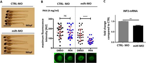
Mitf knockdown model and PAN treatment. (A) Phenotype pictures of Tg(l-fabp:eGFP-DBP) zebrafish backcrossed on AB background that were injected with either a mitf MO or a scrambled MO in a concentration of 250 µM in one to four-cell stage. Pictures were taken at 96 hpf. (B) Tg(l-fabp:eGFP-DBP) zebrafish backcrossed on AB background were injected with a mitf MO or a scrambled MO in a concentration of 250 µM in one to four-cell stage. At 46 hpf fish were treated either with PAN (4 mg/ml) or DMSO. Intensity of circulating eGFP-DBP (arbitrary units) in the retinal vessel plexus of the fish eye was measured at 96 hpf. Representative images of the fluorescence of circulating eGFP-DBP in the retinal vessel plexus of the fish eye 96 hpf are shown at the bottom. n=25–28 per group. (C) Q-PCR for inf2 mRNA expression in whole zebrafish at 120 hpf. Zebrafish (AB strain) were injected with CTRL-MO or a mitf-MO at one to four-cell stage. Data from three different experiments. Significance was tested with two-way ANOVA: **P<0.01, ****P<0.001, ns, non-significant. Scale bars: 500 µm. |