
|
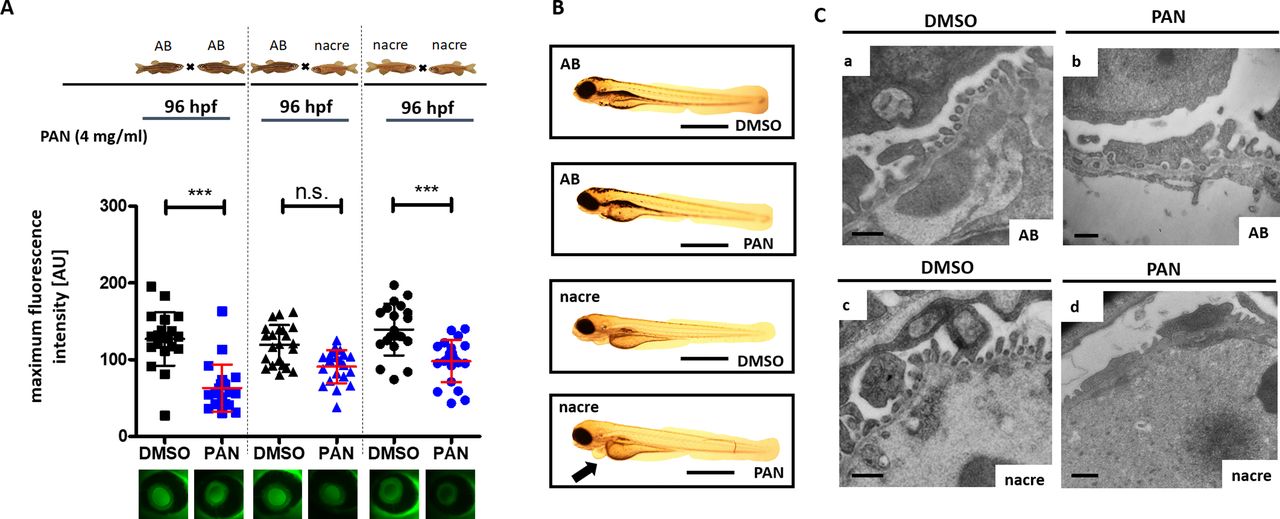
Fig. 1
Influence of genetic background on the proteinuria phenotype. (A) Tg(l-fabp:eGFP-DBP) zebrafish backcrossed on homozygous for either the AB (AB fish) or the nacre (nacw2) background (nacre fish) as well as heterozygous zebrafish for nacre and AB background were treated with PAN at 46 h post-fertilization (hpf). Graph depicts max. fluorescence of circulating eGFP-DBP in the retinal vessel plexus of the fish eye 96 hpf. (B) Representative images of phenotypes of Tg(l-fabp:eGFP-DBP) zebrafish backcrossed on homozygous for either the AB (AB fish) or the nacre (nacw2) background (nacre fish) were treated with PAN (4 mg/ml) or DMSO at 46 hpf as indicated. Pictures were taken at 96 hpf. (C) Representative transmission electron microscopy images of the glomerular filtration barrier of Tg(l-fabp:eGFP-DBP) zebrafish backcrossed on homozygous for either the AB or the nacre (nacw2) background were treated with PAN (4 mg/ml) or DMSO at 46 hpf as indicated. Fish were collected at 120 hpf. Podocyte effacement after PAN treatment was more severe in nacre fish compared to AB fish. ***P<0.001, n.s., non-significant. Scale bars: (B) 500 μm, (C) 500 nm.