
- Title
-
Comparison of efficacy and toxicity of bevacizumab, endostar and apatinib in transgenic and human lung cancer xenograft zebrafish model
- Authors
- Jin, Y., Wei, L., Jiang, Q., Song, X., Teng, C., Fan, C., Lv, Y., Liu, Y., Shen, W., Li, L., Huang, D., Xin, T.
- Source
- Full text @ Sci. Rep.

Anti-angiogenic effects after treatment with angiogenesis inhibitors. (A) Lateral view of subintestinal vessels (SIVs) in fluorescent transgenic zebrafish embryos at 3dpf that were treated with anti-angiogenic drugs at different concentrations for 24 hours. (B) Total area of SIVs (in pixel). (C) The inhibitory rate of SIVs angiogenesis is calculated as difference between total area of SIVs in treated and control groups expressed as percentage of total area of SIVs in control zebrafish embryos. The error bars represent ± SEM. P values were determined by one-way ANOVA followed by Dunnett’s test for multiple comparisons to control. ***p < 0.001 indicate statistically significant difference. SEM, standard error of the mean. |
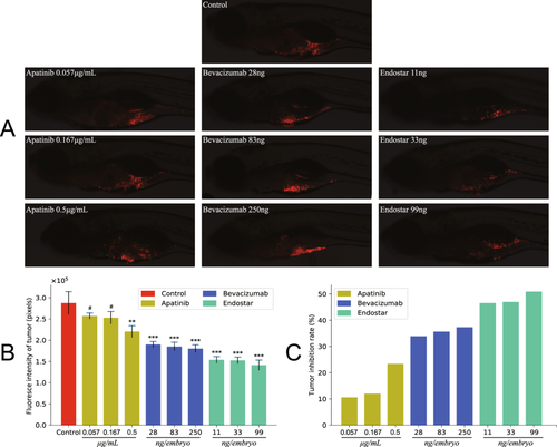
Antitumor effect of anti-angiogenic drugs in the zebrafish embryos. Tumor inhibition effect of three anti-angiogenic drugs in an A549 xenograft zebrafish model. Each compound was evaluated at three different concentrations: bevacizumab (28 ng, 83 ng and 250 ng per embryo), apatinib (0.057 μg/mL, 0.167 μg/mL and 0.5 μg/mL) and endostar (11 ng, 33 ng and 99 ng per embryo). (A) Lateral views of CM-Dil stained A549 cells in 5dpf zebrafish embryos treated with compound for 72 hours. (B) Fluorescence intensity values of A549 cells in zebrafish larvae. (C) Tumor growth inhibition rates is calculated as difference between fluorescence intensities in treated and control groups expressed as percentage of tumor fluorescence intensities in control zebrafish. All quantification data are presented as means ± SEM. P values were calculated with one-way ANOVA followed by Dunnett’s test, **p < 0.01, ***p < 0.001, #p > 0.05 compared to control. |
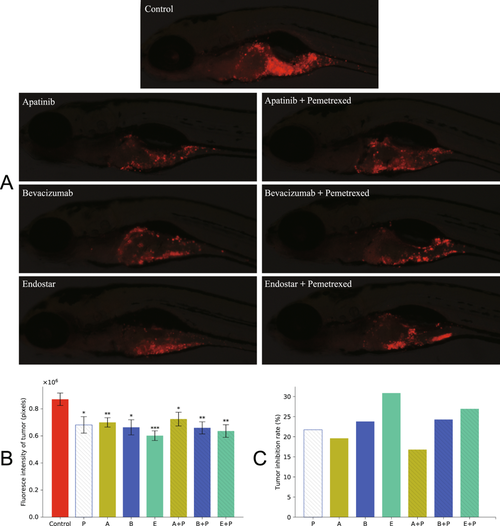
Antitumor activity of anti-angiogenesis alone and in combination with pemetrexed. On 2dpf, CM-Dil labeled A549 xenograft zebrafish embryos were treated with three anti-angiogenesis alone and in combination with pemetrexed for 3 days. Pemetrexed (P) (2 ng per embryo), bevacizumab (B) (28 ng per embryo) and endostar (E) (11 ng per embryo) were injected into embryos and apatinib (A) (0.5 μg/mL) was dissolved in breeding water. (A) Fluorescent images showing A549 xenograft zebrafish larvae at 5dpf treated with different inhibitors. (B) Quantification of fluorescence intensity of tumor. (C) Tumor growth inhibition rates were calculated according to the fluorescence intensities of tumor. Each bar represents the mean ± SEM. One-way ANOVA followed by Dunnett’s test, *p < 0.05, **p < 0.01, ***p < 0.001 when compared with control group. |

Gross morphological changes in the zebrafish larvae following anti-angiogenic compound exposure at 120 hpf. Wild-type zebrafish were collected at 6 hpf and were treated with bevacizumab (28 ng, 83 ng and 250 ng per embryo), endostar (11 ng, 33 ng and 99 ng per embryo) and apatinib (0.22 μg/mL, 0.67 μg/mL and 2 μg/mL dissolved in breeding water) at 28 °C until 120 hpf. Apatinib (0.67 µg/mL) treatment: pericardial edema (arrow); apatinib (2 µg/mL) treatment: pericardial edema (arrow), decreased eye size (arrowhead) and shortened body axis. |