
- Title
-
Absence of histopathological changes in the retina of zebrafish treated with sodium iodate
- Authors
- Sadamoto, K., Yamagiwa, Y., Sakaki, H., Kurata, M.
- Source
- Full text @ J. Vet. Med. Sci.

Representative images of H&E and PCNA staining of larval zebrafish eyes after sodium iodate exposure at 0.1, 0.3 and 1.0 mM, respectively, from 3 to 8 dpf. No significant differences were observed in H&E and PCNA staining between the control group and the treatment groups. PCNA-positive cells were observed in the CMZ and the ONL. Green: PCNA; Blue: DAPI; CMZ: ciliary marginal zone; GCL: ganglion cell layer; INL: inner nuclear layer; ONL: outer nuclear layer. Scale bars indicate 100 µm. |

Representative images of H&E and PCNA staining of juvenile zebrafish retinas after sodium iodate exposure at 0.1, 0.3 and 1.0 mM, respectively, for 7 days. No significant differences were observed in H&E and PCNA staining between the control group and the treatment groups. PCNA-positive cells were observed in the CMZ and the ONL. Green: PCNA; Blue: DAPI; CMZ: ciliary marginal zone; GCL: ganglion cell layer; INL: inner nuclear layer; ONL: outer nuclear layer. Scale bars indicate 100 µm. |
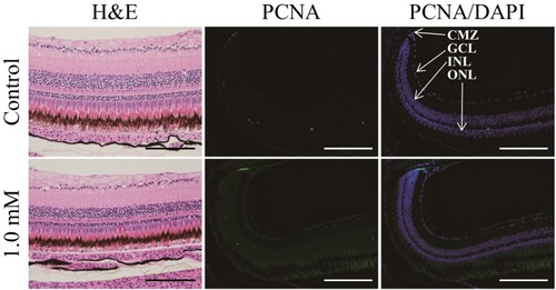
Representative images of H&E and PCNA staining of adult zebrafish retinas after sodium iodate exposure at 1.0 mM for 7 days. No significant differences were observed in H&E and PCNA staining between the control group and the treatment group. PCNA-positive cells were observed in the CMZ and the ONL. Green: PCNA; Blue: DAPI; CMZ: ciliary marginal zone; GCL: ganglion cell layer; INL: inner nuclear layer; ONL: outer nuclear layer. Scale bars indicate 100 µm. |
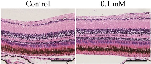
Representative images of H&E staining of adult zebrafish retinas after sodium iodate exposure at 0.1 mM for 30 days. No significant differences were observed between the control group and the treatment group. Scale bars indicate 100 µm. |
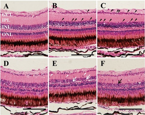
Representative images of H&E staining of adult zebrafish retinas after MNU exposure. Each group of zebrafish was exposed to MNU for 60 min and thereafter maintained under standard conditions for 6 hr, 24 hr, 3 days, 5 days and 8 days, respectively. (A) Vehicle control; (B) Six hr of maintenance after exposure, the pyknotic cells (black arrowheads) started to appear in the INL and the GCL; (C) Twenty four hr of maintenance after exposure, the number of pyknotic cells increased (black arrowheads); (D) Three days of maintenance after exposure, obscuration of retinal structure between the INL and the ONL was observed; (E) Five days of maintenance after exposure, some parts of the INL were fused with the ONL (white arrow); (F) Eight days of maintenance after exposure, accumulation of cell clusters was observed in the INL (black arrow) and cells appeared in the INL (white arrowheads). GCL: ganglion cell layer; IPL: inner plexiform layer; INL: inner nuclear layer; ONL: outer nuclear layer. Scale bars indicate 100 µm. |