
Image
|
Figure Caption
Fig. 1
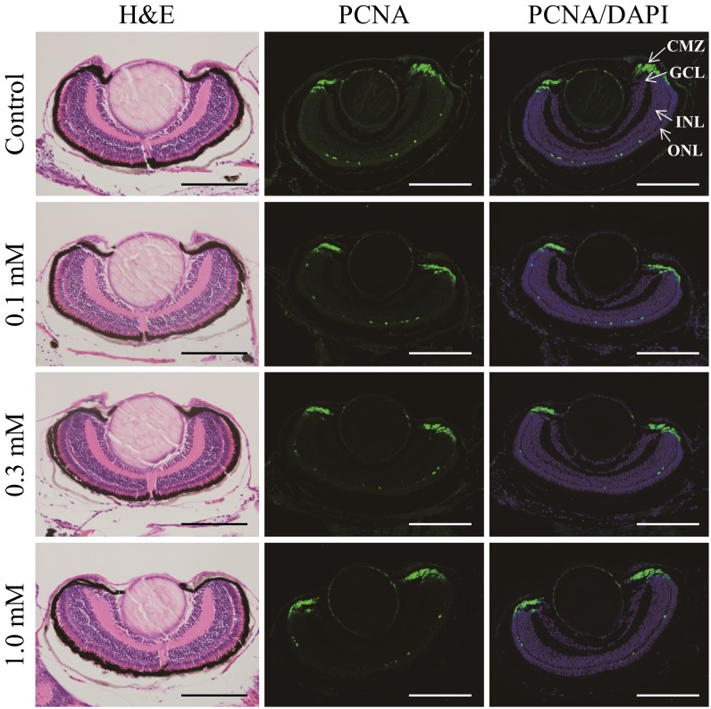
Representative images of H&E and PCNA staining of larval zebrafish eyes after sodium iodate exposure at 0.1, 0.3 and 1.0 mM, respectively, from 3 to 8 dpf. No significant differences were observed in H&E and PCNA staining between the control group and the treatment groups. PCNA-positive cells were observed in the CMZ and the ONL. Green: PCNA; Blue: DAPI; CMZ: ciliary marginal zone; GCL: ganglion cell layer; INL: inner nuclear layer; ONL: outer nuclear layer. Scale bars indicate 100 µm.
Acknowledgments
This image is the copyrighted work of the attributed author or publisher, and
ZFIN has permission only to display this image to its users.
Additional permissions should be obtained from the applicable author or publisher of the image.
Full text @ J. Vet. Med. Sci.