
Fig. 5
- ID
- ZDB-FIG-180810-6
- Publication
- Sadamoto et al., 2018 - Absence of histopathological changes in the retina of zebrafish treated with sodium iodate
- Other Figures
- All Figure Page
- Back to All Figure Page
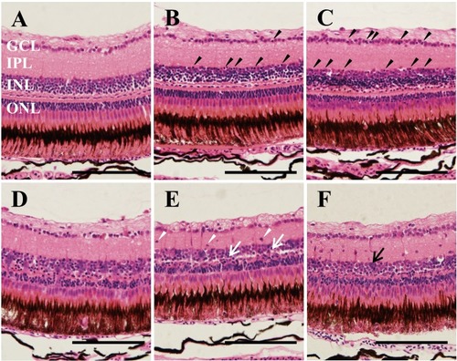
Representative images of H&E staining of adult zebrafish retinas after MNU exposure. Each group of zebrafish was exposed to MNU for 60 min and thereafter maintained under standard conditions for 6 hr, 24 hr, 3 days, 5 days and 8 days, respectively. (A) Vehicle control; (B) Six hr of maintenance after exposure, the pyknotic cells (black arrowheads) started to appear in the INL and the GCL; (C) Twenty four hr of maintenance after exposure, the number of pyknotic cells increased (black arrowheads); (D) Three days of maintenance after exposure, obscuration of retinal structure between the INL and the ONL was observed; (E) Five days of maintenance after exposure, some parts of the INL were fused with the ONL (white arrow); (F) Eight days of maintenance after exposure, accumulation of cell clusters was observed in the INL (black arrow) and cells appeared in the INL (white arrowheads). GCL: ganglion cell layer; IPL: inner plexiform layer; INL: inner nuclear layer; ONL: outer nuclear layer. Scale bars indicate 100 µm. |