
- Title
-
Irisin regulates cardiac physiology in zebrafish
- Authors
- Sundarrajan, L., Yeung, C., Hahn, L., Weber, L.P., Unniappan, S.
- Source
- Full text @ PLoS One

ZFIN is incorporating published figure images and captions as part of an ongoing project. Figures from some publications have not yet been curated, or are not available for display because of copyright restrictions. EXPRESSION / LABELING:
|
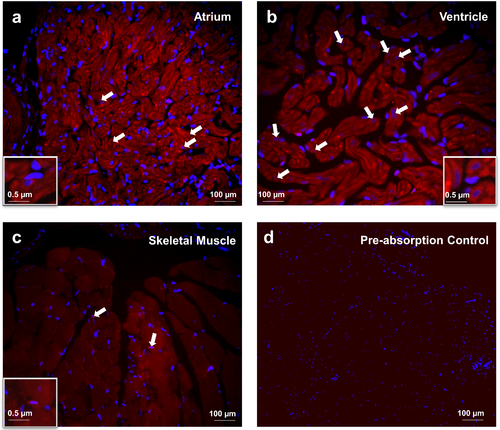
Irisin immunoreactivity was detected in the atrium (A) and ventricle (B) of heart and (C) skeletal muscle tissues of zebrafish. Irisin immunoreactivity was detected in atrial and ventricular cardiomyocytes of zebrafish (A,B) In skeletal muscle, irisin immunoreactivity (Catalog# ab131390, 1:3000, Abcam, Ramona, Massachusetts) was detected at the myofibril filament within the myotubule (C). Preabsorption control of irisin was used as negative control (D). Nuclei are stained blue (DAPI). Images were taken at 40X magnification and scale bar = 100 μm (and 0.5 μm for inset). EXPRESSION / LABELING:
|
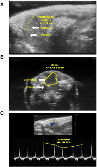
Representative long-axis (A), short axis (B) brightness mode (B-mode) view of adult zebrafish heart. Zebrafish were anesthetized, imaged and the long axis mode was considered to calculate the ventricular length (A), while short axis was considered to calculate ventricular area (B) (represented by blue line). Blood flow from atrium to ventricle through the atrioventricular valve are indicated by color (blue) in panel C using color flow Doppler mode and heart rate measurements were calculated by number of heart beats per 10 s during the B-mode ultrasound video loop, and converted to beats per minutes (bpm). |