
FIGURE
Fig. 2
- ID
- ZDB-FIG-180122-30
- Publication
- Sundarrajan et al., 2017 - Irisin regulates cardiac physiology in zebrafish
- Other Figures
- All Figure Page
- Back to All Figure Page
Fig. 2
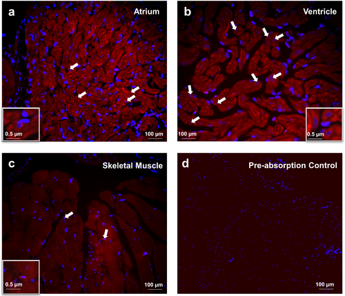
Irisin immunoreactivity was detected in the atrium (A) and ventricle (B) of heart and (C) skeletal muscle tissues of zebrafish. Irisin immunoreactivity was detected in atrial and ventricular cardiomyocytes of zebrafish (A,B) In skeletal muscle, irisin immunoreactivity (Catalog# ab131390, 1:3000, Abcam, Ramona, Massachusetts) was detected at the myofibril filament within the myotubule (C). Preabsorption control of irisin was used as negative control (D). Nuclei are stained blue (DAPI). Images were taken at 40X magnification and scale bar = 100 μm (and 0.5 μm for inset). |
Expression Data
| Antibody: | |
|---|---|
| Fish: | |
| Anatomical Terms: | |
| Stage: | Days 45-89 |
Expression Detail
Antibody Labeling
Phenotype Data
Phenotype Detail
Acknowledgments
This image is the copyrighted work of the attributed author or publisher, and
ZFIN has permission only to display this image to its users.
Additional permissions should be obtained from the applicable author or publisher of the image.
Full text @ PLoS One