
- Title
-
The Effect of Retinal Melanin on Optical Coherence Tomography Images
- Authors
- Wilk, M.A., Huckenpahler, A.L., Collery, R.F., Link, B.A., Carroll, J.
- Source
- Full text @ Transl Vis Sci Technol
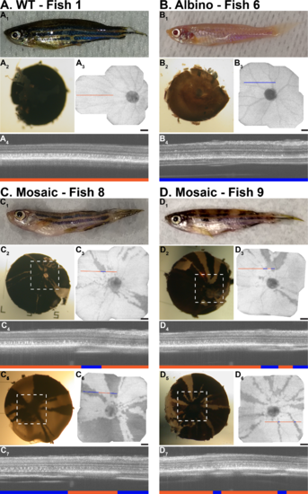
Pigmentation patterns of zebrafish strains. (A) WT zebrafish. (A1) External pigmentation of normal zebrafish. (A2) Ex vivo, posterior view of the normal eyecup. (A3) En face OCT image from the same retina as (A2). (A4) In vivo cross-section of the retina at the location indicated by the orange line in (A3). (B) Albino zebrafish. (B1) External pigmentation of albino zebrafish. (B2) Ex vivo, posterior view of the albino eyecup. (B3) En face OCT image from the same retina as (B2). (B4) In vivo cross-section of the retina at the location indicated by the blue line in (B3). (C–D) Two examples of mosaic zebrafish with unique pigmentation patterns. (C1, D1) External pigmentation of mosaic zebrafish. Retinal images from the right eyes (C2–C4, D2–D4) and left eyes (C5–C7, D5–D7) of two mosaic zebrafish. Posterior eyecups and en face OCT images show identical pigmentation patterns. Dashed white boxes on the eyecup denote the approximate location of the corresponding en face OCT images. For all fish, lines overlaid on the en face OCT mark the location of the B-scans below. Orange lines mark locations of melanin pigment while blue lines denote no pigment. Areas of pigment display bright RPE bands while areas lacking pigment have reduced RPE reflectance. The choroid and sclera are more visible in areas lacking pigment. Scale bars: 100 μm. All B-scans are 475 μm wide and tall and are displayed in the logarithmic format. PHENOTYPE:
|

Differences in LRP peaks with pigmentation in zebrafish. (A) Wild type fish with consistent RPE reflectance and width. (B) Albino fish, whose RPE is distinctly dimmer than the WT. (C) Mosaic fish, where the left LRP was generated in an area with melanin pigment and the right LRP was generated in an area lacking pigment. When melanin in present, the RPE band is wider and has increased reflectance. When melanin is absent, the sclera also is visible posterior to the RPE and choroid. Wild type and albino images are 225 μm wide. Mosaic fish image is 475 μm wide. All images are 479 μm tall. Images are displayed in linear format with intensity values normalized to stretch between 0 and 255 for display only. RNFL, retinal nerve fiber layer; IPL, inner plexiform layer; OPL, outer plexiform layer; PR, photoreceptor layers. PHENOTYPE:
|
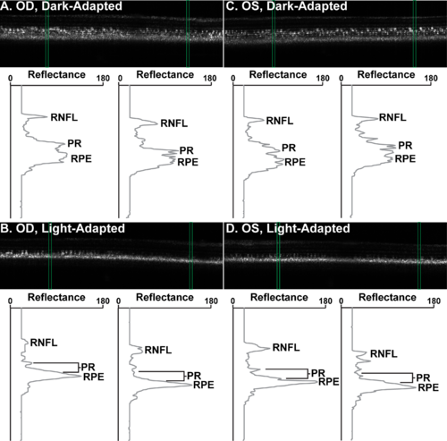
Light- and dark-adapted zebrafish OCT. Green boxes over images denote the location of the LRPs below. Right eye ([A]; OD) and left eye ([C]; OS) of WT zebrafish under dark adaptation. The RPE appears grainy and dispersed. Photoreceptor layers are distinct. (B, D) Corresponding light-adapted retinas. The RPE is more condensed and reflective. The RPE appears closer to the photoreceptor layers, which are much less distinct. Images are 475 μm wide and 479 μm tall. Images are displayed in linear format with intensity values normalized to stretch between 0 and 255 for display only. |