
FIGURE
Fig. 6
- ID
- ZDB-FIG-180104-16
- Publication
- Wilk et al., 2017 - The Effect of Retinal Melanin on Optical Coherence Tomography Images
- Other Figures
- All Figure Page
- Back to All Figure Page
Fig. 6
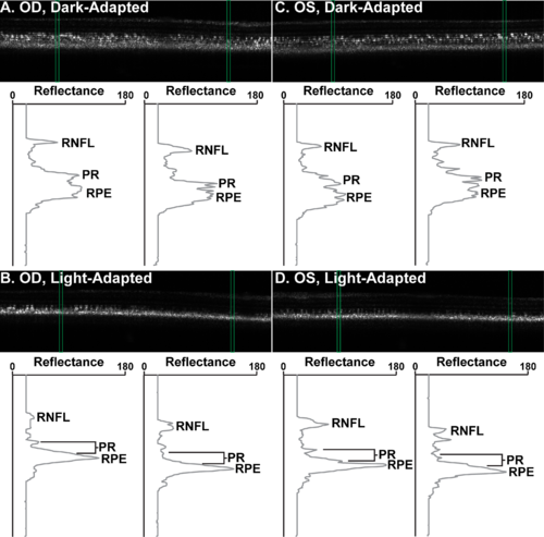
Light- and dark-adapted zebrafish OCT. Green boxes over images denote the location of the LRPs below. Right eye ([A]; OD) and left eye ([C]; OS) of WT zebrafish under dark adaptation. The RPE appears grainy and dispersed. Photoreceptor layers are distinct. (B, D) Corresponding light-adapted retinas. The RPE is more condensed and reflective. The RPE appears closer to the photoreceptor layers, which are much less distinct. Images are 475 μm wide and 479 μm tall. Images are displayed in linear format with intensity values normalized to stretch between 0 and 255 for display only. |
Expression Data
Expression Detail
Antibody Labeling
Phenotype Data
Phenotype Detail
Acknowledgments
This image is the copyrighted work of the attributed author or publisher, and
ZFIN has permission only to display this image to its users.
Additional permissions should be obtained from the applicable author or publisher of the image.
Full text @ Transl Vis Sci Technol