
- Title
-
Short-term overfeeding of zebrafish with normal or high-fat diet as a model for the development of metabolically healthy versus unhealthy obesity
- Authors
- Landgraf, K., Schuster, S., Meusel, A., Garten, A., Riemer, T., Schleinitz, D., Kiess, W., Körner, A.
- Source
- Full text @ BMC Physiol.
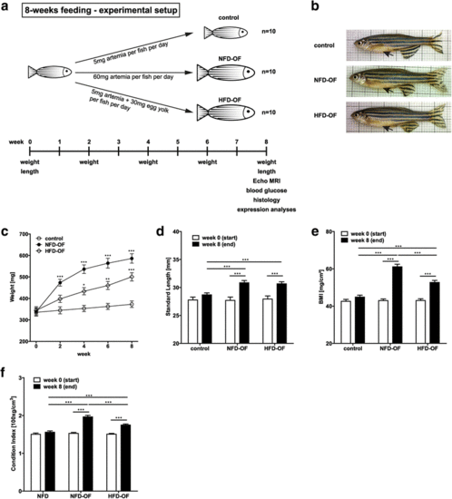
Overfeeding of adult zebrafish with either NFD or HFD leads to an obese phenotype. a Schematic overview of the 8-week feeding protocol and phenotyping of zebrafish. b Exemplary images of zebrafish included in each of the analyzed dietary groups at week 8 of feeding. Black arrows point to the abdominal region. Overfeeding of zebrafish with either NFD or HFD resulted in a significant increase in weight (c), standard length (d), BMI (e), and Fulton’s condition index (f) compared to control zebrafish. Statistical analyses were performed using Two-Way ANOVA and Bonferroni post-test and significant p-values are indicated. *, p < 0.05; **, p < 0.01; ***, p < 0.001; NFD, normal fat diet; HFD, high-fat-diet; OF, overfeeding; BMI, body mass index; MRI, Magnetic Resonance Imaging; AT, adipose tissue PHENOTYPE:
|
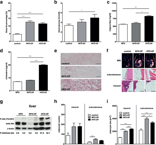
Phenotypic characterization of diet-induced obesity in zebrafish. Overfeeding of zebrafish with either NFD or HFD resulted in a significant increase in body fat percentage (a) compared to control zebrafish. HFD-OF but not NFD-OF zebrafish showed significantly elevated fasting blood glucose levels (b), triglyceride levels (c) and cholesterol levels (d), and a prominent accumulation of lipids in the liver as indicated by Oilred-O staining of liver sections (e). Fat distribution was analyzed by MRI and hematoxylin-eosin staining of zebrafish cross sections and revealed an increase in the amount of both subcutaneous and visceral AT in NFD-OF and HFD-OF compared to control zebrafish (f). Exemplary images of zebrafish included in each of the analyzed dietary groups at week 8 of feeding are shown. Asterisk indicates intramuscular fat deposition in HFD-OF fish. Immunoblot analyses of liver lysates showed increased phosphorylation of Akt at Thr307 (Thr308 in human) in HFD-OF compared to NFD-OF and control zebrafish. The ratio of P-Akt to total Akt was analyzed using ImageJ software and is given underneath the immunoblot images (g). Detection of β-Actin served as loading control. Visceral and subcutaneous adipocyte number and adipocyte size were determined from hematoxylin-eosin-stained zebrafish sections using ImageJ software and were increased after overfeeding with NFD or HFD (h, i). Statistical analyses were performed using One-Way ANOVA and Bonferroni post-test and significant p-values are indicated. *, p < 0.05; **, p < 0.01; ***, p < 0.001; Scale bar in (h) represents 50 μm, Scale bar in (i) represents 100 μm; NFD, normal fat diet; HFD, high-fat-diet; OF, overfeeding; BMI, body mass index; MRI, Magnetic Resonance Imaging; AT, adipose tissue |

ZFIN is incorporating published figure images and captions as part of an ongoing project. Figures from some publications have not yet been curated, or are not available for display because of copyright restrictions. |