- Title
-
Sustainable, Rapid Synthesis of Bright-Luminescent CuInS2-ZnS Alloyed Nanocrystals: Multistage Nano-xenotoxicity Assessment and Intravital Fluorescence Bioimaging in Zebrafish-Embryos
- Authors
- Chetty, S.S., Praneetha, S., Basu, S., Sachidanandan, C., Murugan, A.V.
- Source
- Full text @ Sci. Rep.
|
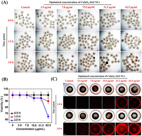
In vivo nano-xenotoxicity assessment in 6 hpf zebrafish embryos.(A) Bright-field microscopic images at three-time points of 6 hpf zebrafish embryos (N = 25) treated with different concentration MUA-functionalized CIZS-NCs for 3.0 h. (B) Embryos viability (%). (C) Bright-field (a,c) with fluorescence (b,d) microscopic images at two-time points indicating relative uptake of MUA-functionalized CIZS-NCs in 6 hpf zebrafish embryos. |
|
In vivo nano-xenotoxicity assessment in 24 hpf zebrafish embryos. (A) Bright-field microscopic images at three-time points of 24 hpf zebrafish embryos (N = 25) treated with different concentration MUA-functionalized CIZS-NCs for 3.0 h. (B) Embryos viability (%). (C) Bright-field (a,c) with fluorescence (b,d) microscopic images at two-time points indicating relative uptake of MUA-functionalized CIZS-NCs in 6 hpf zebrafish embryos. |
|
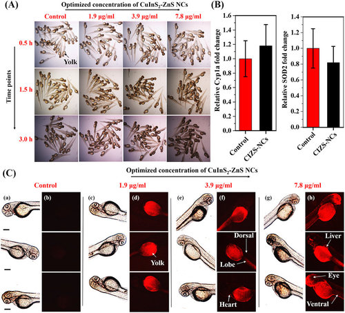
In vivo nano-xenotoxicity assessment and intravital imaging in 3 dpf zebrafish embryos.(A) Bright-field microscopic images of 3 dpf zebrafish embryos (N = 25) treated with optimized concentrations of MUA-functionalized CIZS-NCs for 3.0 h. (B) CYP1A and SOD2 gene expression profile in embryos incubated with 7.8 µg/ml concentration of MUA-functionalized CIZS-NCs. (C) Bright-field (a,c,e,g) and fluorescence (b,d,f,h) microscopic images of 3 dpf zebrafish embryos co-incubated with optimized concentrations of MUA-functionalized CIZS-NCs at 3.0 h. |


