
Image
|
Figure Caption
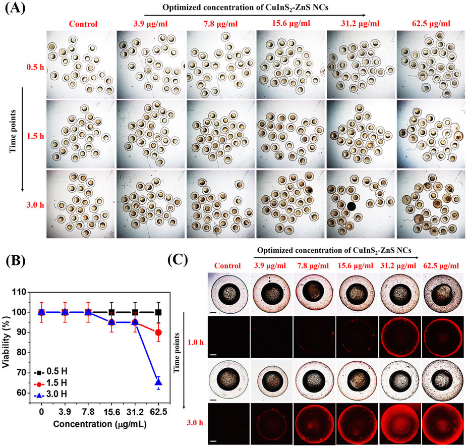
Fig. 8
In vivo nano-xenotoxicity assessment in 6 hpf zebrafish embryos.(A) Bright-field microscopic images at three-time points of 6 hpf zebrafish embryos (N = 25) treated with different concentration MUA-functionalized CIZS-NCs for 3.0 h. (B) Embryos viability (%). (C) Bright-field (a,c) with fluorescence (b,d) microscopic images at two-time points indicating relative uptake of MUA-functionalized CIZS-NCs in 6 hpf zebrafish embryos.
Acknowledgments
This image is the copyrighted work of the attributed author or publisher, and
ZFIN has permission only to display this image to its users.
Additional permissions should be obtained from the applicable author or publisher of the image.
Full text @ Sci. Rep.