
Image
|
Figure Caption
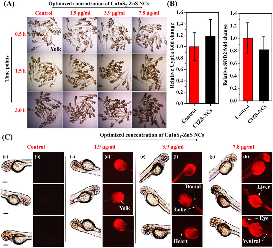
Fig. 10
In vivo nano-xenotoxicity assessment and intravital imaging in 3 dpf zebrafish embryos.(A) Bright-field microscopic images of 3 dpf zebrafish embryos (N = 25) treated with optimized concentrations of MUA-functionalized CIZS-NCs for 3.0 h. (B) CYP1A and SOD2 gene expression profile in embryos incubated with 7.8 µg/ml concentration of MUA-functionalized CIZS-NCs. (C) Bright-field (a,c,e,g) and fluorescence (b,d,f,h) microscopic images of 3 dpf zebrafish embryos co-incubated with optimized concentrations of MUA-functionalized CIZS-NCs at 3.0 h.
Acknowledgments
This image is the copyrighted work of the attributed author or publisher, and
ZFIN has permission only to display this image to its users.
Additional permissions should be obtained from the applicable author or publisher of the image.
Full text @ Sci. Rep.