
- Title
-
Characterization and Expression of the Zebrafish qki Paralogs
- Authors
- Radomska, K.J., Sager, J., Farnsworth, B., Tellgren-Roth, Å., Tuveri, G., Peuckert, C., Kettunen, P., Jazin, E., Emilsson, L.S.
- Source
- Full text @ PLoS One

ZFIN is incorporating published figure images and captions as part of an ongoing project. Figures from some publications have not yet been curated, or are not available for display because of copyright restrictions. |
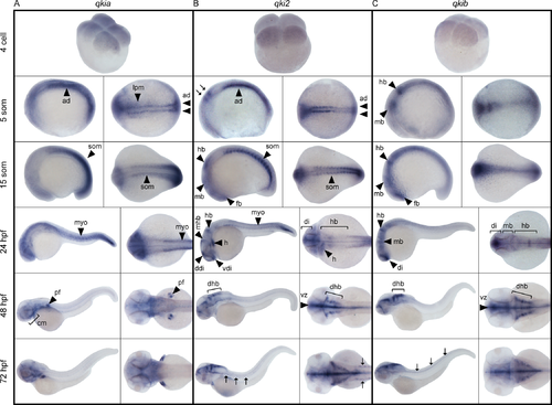
Spatiotemporal expression of qki genes during development. Representative images of whole mount in situ hybridization using probes detecting qkia (A), qki2 (B) and qkib (C) expression. Developmental stages are expressed as the number of cells, somites (som), or hours post-fertilization (hpf). Lateral (left panels) and dorsal (right panels) views are shown, anterior to the left. Arrows in B at the 5 somite stage indicate the hindbrain primordium, while arrows at 72 hpf indicate the presumptive lateral line Schwann cells and in C, developing trunk neural tube. ad: adaxial cells, lpm: lateral paraxial mesoderm, som: somites, fb: forebrain, di: diencephalon, vdi: ventral diencephalon, ddi: dorsal diencephalon, dhb: dorsolateral hindbrain, mb: midbrain, mhb: midbrain/hindbrain boundary, hb: hindbrain, vz: ventricular zone, myo: myotome, pf: pectoral fin, cm: cranial muscles, h: heart. EXPRESSION / LABELING:
|
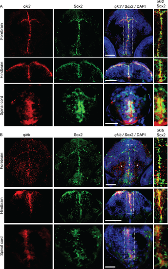
qki2 and qkib are expressed in neural progenitor cells. Representative images of combined in situ hybridization and immunofluorescence detecting qki2 (A) and qkib probes (B), both shown in red. Sox2 antibody is shown in green and DAPI in blue. Images shown are within the forebrain, hindbrain and spinal cord and overlays are denoted in the figure header. Scale bars are 50μm for forebrain and hindbrain images, and 20μm for spinal cord images. Asterisks indicate staining artefacts. EXPRESSION / LABELING:
|
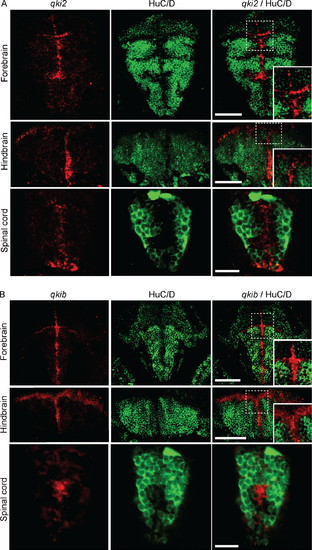
qki2 and qkib are absent from differentiated neurons in developing zebrafish. Representative images of combined in situ hybridization and immunofluorescence detecting qki2 (A) and qkib probes (B), both shown in red. HuC/D antibody is shown in green. Images shown are within the forebrain, hindbrain and spinal cord. EXPRESSION / LABELING:
|