
Fig. 5
- ID
- ZDB-IMAGE-171106-1
- Genes
- Publication
- Radomska et al., 2016 - Characterization and Expression of the Zebrafish qki Paralogs
- All Figures
- Figures for Radomska et al., 2016
|
Fig. 5
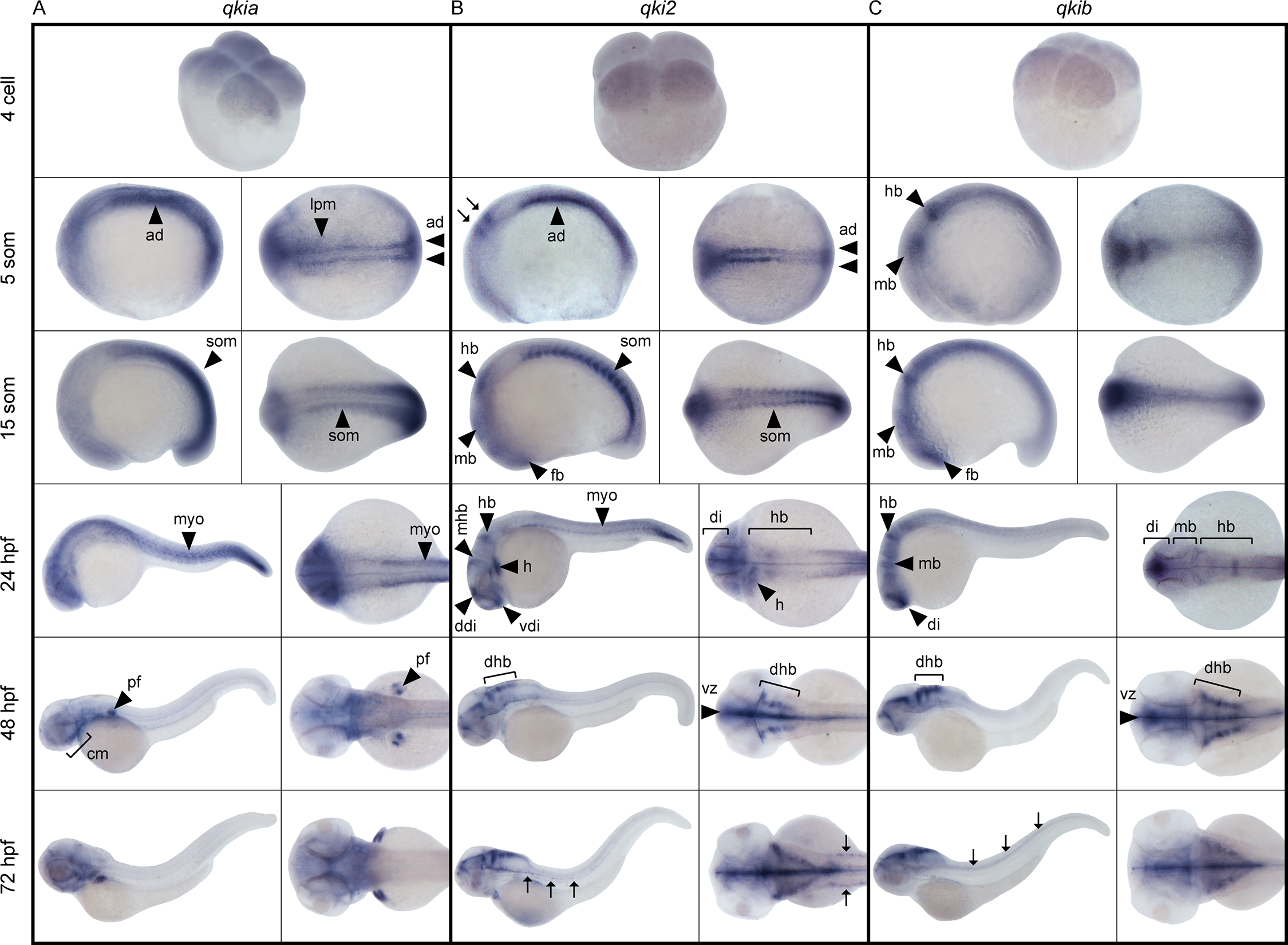
Spatiotemporal expression of qki genes during development.
Representative images of whole mount in situ hybridization using probes detecting qkia (A), qki2 (B) and qkib (C) expression. Developmental stages are expressed as the number of cells, somites (som), or hours post-fertilization (hpf). Lateral (left panels) and dorsal (right panels) views are shown, anterior to the left. Arrows in B at the 5 somite stage indicate the hindbrain primordium, while arrows at 72 hpf indicate the presumptive lateral line Schwann cells and in C, developing trunk neural tube. ad: adaxial cells, lpm: lateral paraxial mesoderm, som: somites, fb: forebrain, di: diencephalon, vdi: ventral diencephalon, ddi: dorsal diencephalon, dhb: dorsolateral hindbrain, mb: midbrain, mhb: midbrain/hindbrain boundary, hb: hindbrain, vz: ventricular zone, myo: myotome, pf: pectoral fin, cm: cranial muscles, h: heart.