
- Title
-
Selective disruption of vascular endothelium of zebrafish embryos by ultrafast laser microsurgical treatment
- Authors
- Woo, S.Y., Moon, H.Y., Kim, T.G., Lee, H.S., Sidhu, M.S., Kim, C., Jeon, J.P., Jeoung, S.C.
- Source
- Full text @ Biomed. Opt. Express
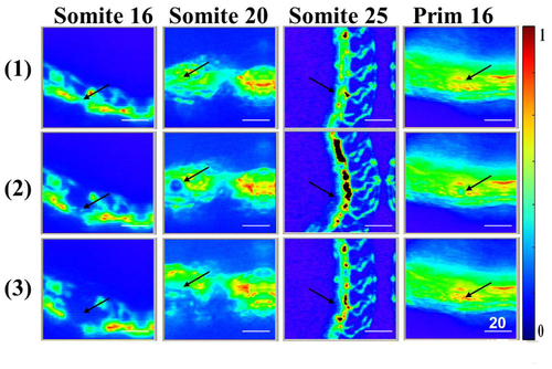
Typical confocal microscopic images (1) before and (2 and 3) after the ultrafast photo-induced disruption of the vascular structure in zebrafish embryos in four different developmental stages. The images were taken with a time lapse of 0.5 sec. The black arrows indicate the targeted area of the ultrafast laser beam. Scale bar: 20 μm. |
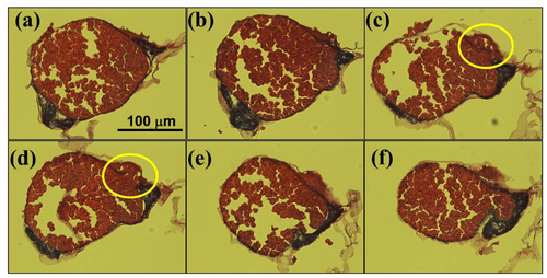
Series of optical images of the zebrafish embryo sections. The images from (a) to (f) were successively obtained from the samples sectioned by crytome with a thickness of 20 μm. Only the middle sections (c-d) exhibit complete disruption marked with yellow circles of the angiogenesis in addition to the feature of yolk cell extrusion. However, Figs. 3(a)-3(b) and 3(e)-3(f) reveal only the extruded yolk cells from the embryos and they maintain intact vascular structure parts at the border. Scale bar: 100 μm. |
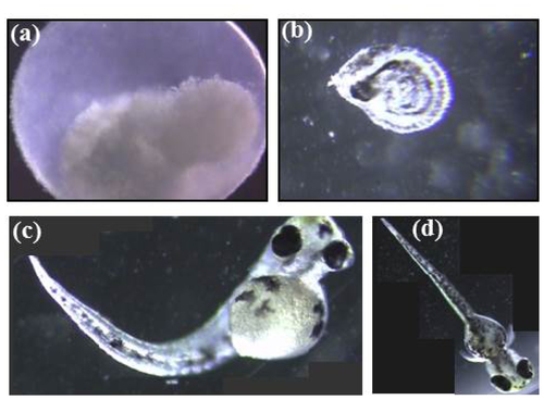
The typical final status for each development stage of the laser-treated zebrafish embryos. (a) embryo did not progress further in development after the laser irradiation; (b) embryo developed but did not hatch and eventually died; (c) embryo hatched but the juvenile fish did not have a good shape; and (d) embryo finished its developmental process, hatched well, and grew normally even though the embryo’s vascular structure was treated with an ultrafast laser. |