
Image
|
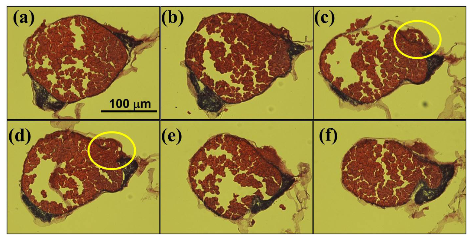
Figure Caption
Fig. 3
Series of optical images of the zebrafish embryo sections. The images from (a) to (f) were successively obtained from the samples sectioned by crytome with a thickness of 20 μm. Only the middle sections (c-d) exhibit complete disruption marked with yellow circles of the angiogenesis in addition to the feature of yolk cell extrusion. However, Figs. 3(a)-3(b) and 3(e)-3(f) reveal only the extruded yolk cells from the embryos and they maintain intact vascular structure parts at the border. Scale bar: 100 μm.
Acknowledgments
This image is the copyrighted work of the attributed author or publisher, and
ZFIN has permission only to display this image to its users.
Additional permissions should be obtained from the applicable author or publisher of the image.
Full text @ Biomed. Opt. Express