
Image
|
Figure Caption
Fig. 2
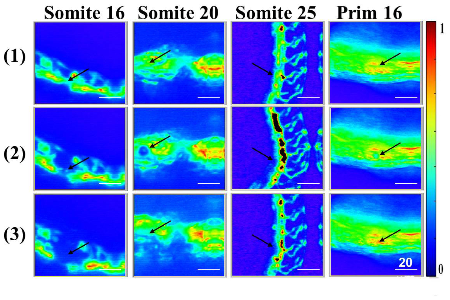
Typical confocal microscopic images (1) before and (2 and 3) after the ultrafast photo-induced disruption of the vascular structure in zebrafish embryos in four different developmental stages. The images were taken with a time lapse of 0.5 sec. The black arrows indicate the targeted area of the ultrafast laser beam. Scale bar: 20 μm.
Acknowledgments
This image is the copyrighted work of the attributed author or publisher, and
ZFIN has permission only to display this image to its users.
Additional permissions should be obtained from the applicable author or publisher of the image.
Full text @ Biomed. Opt. Express