- Title
-
Identification and expression of Lypc, a novel dark-inducible member of Ly6 superfamily in zebrafish Danio rerio
- Authors
- Li, L., Ji, D., Teng, L., Zhang, S., Li, H.
- Source
- Full text @ Gene
|
The expression of lypc by qRT-PCR: (A) Expression profile of lypc at different tissues of adult zebrafish as measured by Real-Time PCR. The mRNA expressions of lypc and β-actin were detected at skin, heart, muscle, gill, liver, intestine, brain, eyes. (B) Expression profile of lypc at different stages of embryonic development as measured by Real-Time PCR. The mRNA expressions of lypc and β-actin were detected at 0 hpf, 2.25 hpf, 4 hpf, 6 hpf, 10 hpf, 14 hpf, 24 hpf, 48 hpf, 72 hpf, 96 hpf. Fold difference was calculated as 2- ΔΔCt with zebrafish β-actin as a reference gene. Vertical bars represent the mean ± SEM (n = 3). |
|
The expression pattern of lypc in early development: (A): Long-pec embryo (48 hpf), pigment cells reach terminal differentiation along dorsal strip (arrows) (B): pigment cells in the retina of embryo (48 hpf) (C): pigment cells in tail (48 hpf). (D–F): By 72 h, lypc strongly expresses on ventral side of yolk sac. Lypc positive cells have organized along the dorsal, ventral and yolk stripes. Red arrow: dorsal stripe, ventral yolk stripes (G–H). Lateral view of larva of 96 hpf. The expression pattern of lypc was mainly restricted to the ventral yolk stripes. (I–J) The longitudinal section embryos at 72 hpf after in situ hybridization (K) is showing the traverse section of embryo at 72 hpf. Strong signal in epidermal cells in the trunk and tail reveals that lypc expresses in pigment cells. (L) The retina of zebrafish embryo at 72 hpf. |
|
Ocular transverse section after WISH: (A) In situ hybridization for lypc in 4dpf zebrafish larvae eye. The signal notes that expression in the retinal pigment cell layer. (B) Further magnification of the eye shows prominent expression of lypc in RPE, retinal pigment epithelium. (C) Microstructure of RPE. ONL, outer nuclear layer. EXPRESSION / LABELING:
|
|
The expression of lypc in casper mutant. (A) 2 dpf (B) 3 dpf (C) 4 dpf (D) 5 dpf wild-type zebrafish embryo (left), embryos of casper mutant (right). |
|
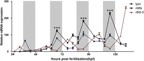
Circadian expression of lypc. Expression analysis was performed on larvae raised on an LD cycle until 5 dpf. White and gray backgrounds represent light and dark phases. Mitfa is used as a positive control and nfil3-5 as a negative control. Statistically significant differences between the expression peak and through on each day (Fisher′s T-test) are indicated:*P < 0.05, **P < 0.01, ***P < 0.001. Error bars indicate s.e.m. |
Reprinted from Gene, 574(1), Li, L., Ji, D., Teng, L., Zhang, S., Li, H., Identification and expression of Lypc, a novel dark-inducible member of Ly6 superfamily in zebrafish Danio rerio, 69-75, Copyright (2015) with permission from Elsevier. Full text @ Gene


