- Title
-
Recoverin depletion accelerates cone photoresponse recovery
- Authors
- Zang, J., Keim, J., Kastenhuber, E., Gesemann, M., Neuhauss, S.C.
- Source
- Full text @ Open Biol.
|
Expression of rcv genes in 3 dpf and 5 dpf zebrafish larvae. (a–h) All the rcv genes except rcv1b showed expression in 3 dpf larvae retina. All the rcv genes were expressed in the pineal gland. (i–l) rcv1b still showed no expression in the 5 dpf larval retina. Scale bar (=50 µm) applies to all panels. |
|
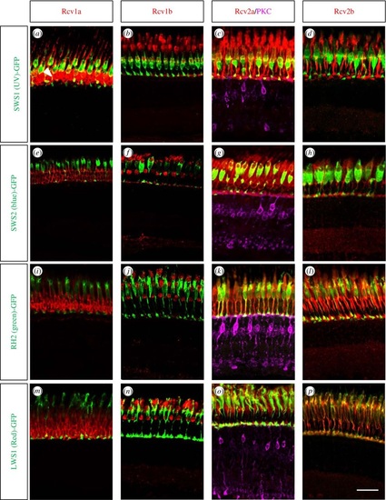
Zebrafish Recoverins are expressed in the different cone subtypes. Adult retinal sections from transgenic zebrafish highlighting the different cone subtypes were co-stained with Rcv antibodies. White arrowhead in (a) marked the rod photoreceptors. While Rcv1b, Rcv2a and Rcv2b proteins are present in all cone subtypes, Rcv1a is only expressed in rods and UV cones. In order to highlight also ON-bipolar cells, Rcv2a antibodies were supplemented with PKC antibodies (violet staining). Scale bar (=20 µm) applies to all panels. EXPRESSION / LABELING:
|
|
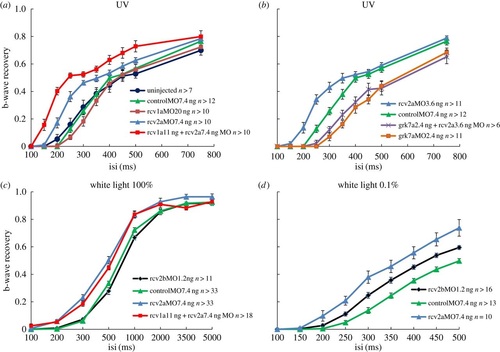
Cone photoresponse recovery is accelerated in Rcv-deficient larvae. Time course of b-wave recovery in (a,b) UV spectral ERG, (c) maximum white light ERG and (d) dim white light ERG are shown. Under UV light conditions recoverin2a and combination of recoverin1a and recoverin2a are effective in accelerating the response recovery (a), whereas depleting the effort kinase Grk7a as expected prolongs the recovery time (b). Note that Rcv2a knockdown accelerates response recovery under normal and dim white light conditions, whereas Rcv2b only accelerates recovery under dim light conditions (c,d). Data are presented as mean ± s.e.m. |
|
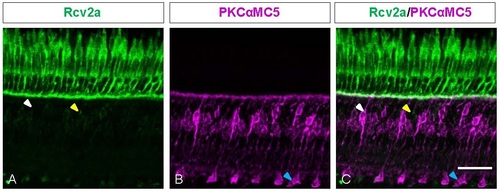
Co-staining of Rcv2a and PKC Antibodies on Retina Sections. Z-projections of confocal image stacks of immunochemical staining on adult retinas. White arrowhead marked the bipolar cell which was labeled by both Rcv2a and PKC antibodies. Yellow arrowhead marked the bipolar cell which was only labeled by Rcv2a antibody. Blue arrowhead marked the ON-bipolar cell which was only labeled by PKC antibody. Scale bar=20 µm. EXPRESSION / LABELING:
|


