
- Title
-
Cardiac Development in Zebrafish and Human Embryonic Stem Cells Is Inhibited by Exposure to Tobacco Cigarettes and E-Cigarettes
- Authors
- Palpant, N.J., Hofsteen, P., Pabon, L., Reinecke, H., Murry, C.E.
- Source
- Full text @ PLoS One
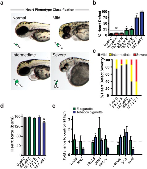
Cardiac developmental defects observed in zebrafish treated with cigarette smoke. (a) Representative whole mount images of zebrafish at 72 hpe showing normal, mild, intermediate, and severe cardiac developmental defects. v = ventricle, At = atrium. (b-c) Analysis of percent zebrafish with heart defects (b) severity of heart defects and (c). (d) Analysis of heart function in control, e-cigarette and tobacco treated groups at 72 hpe. (e) Quantitative RT-PCR analysis (fold change from control) of a panel of genes with critical roles in early heart development at 24 hpe. n ≥ 3 (independent experiments with each n containing between 24?48 animals per treatment). For qRT-PCR, n = 3 with each n consisting of 28?35 embryos from independent breeding pools. * P < 0.05, hpe = hours post exposure; N = Nicotine, E = E-cigarette, T = Tobacco. |
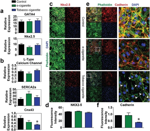
Analysis of hESC derived fetal cardiomyocyte transcription factor, calcium handling, and junction protein expression. (a) Expression level of cardiac transcription factors GATA4 and NKX2.5 (a) and calcium handling proteins including the L-type calcium channel and SERCA2a, and the junctional protein CNX43 (b) by quantitative RT-PCR in cells treated with 6.8 �M e-cigarette or tobacco cigarette extracts vs. control. (c-d) Representative immunocytochemistry (c) and quantification (e) for NKX2.5 in fetal cardiomyocytes with various cigarette treatments compared to control. (e-f) Representative immunohistochemistry (e) and quantification (f) for the junction protein cadherin in fetal hESC cardiomyocytes with various cigarette treatments compared to control. Inset shown to the right. Arrows indicate perinuclear expression of cadherin. n ≥ 6 per group. Scale bar = 100 �m. * P < 0.05. |

Analysis of cardiac myofilament and structural protein expression. (a) Quantitative RT-PCR analysis of early developmental myofilament proteins including the atrial myosin light chain MLC2a, the myosin isoform α-MHC and cardiac troponin T (cTnT) in cells treated with 6.8 �M e-cigarette or tobacco cigarette extracts vs. control. (b-c) Immunohistochemistry (b) and quantification (c) of the myofilament proteins cardiac troponin T (cTnT) in combination with phalloidin and nuclear counterstain DAPI in control cells or those treated with 6.8 �M e-cigarette or 6.8 �M tobacco cigarette extracts. Scale bar = 100 �m for cTnT. (d) Quantitative RT-PCR analysis of mature developmental myofilament isoforms including the ventricular myosin light chain MLC2v and the myosin isoform β-MHC in cells treated 6.8 �M e-cigarette or tobacco cigarette extracts vs. control. (e) Quantitation of sarcomere length as measured from samples stained for α-actinin by immunohistochemistry comparing control vs. 6.8 �M e-cigarette or tobacco cigarette. (f-h) Quantitative RT-PCR (f) immunohistochemistry (g) and quantification of IHC (h) for the immature cardiac marker smooth muscle actin (SMA) in control vs. cells treated with 6.8 �M e-cigarette or tobacco cigarette extract. n ≥ 6 per group. Scale bar = 100 �m. * P < 0.05. |