
- Title
-
A Missense Change in the ATG4D Gene Links Aberrant Autophagy to a Neurodegenerative Vacuolar Storage Disease
- Authors
- Kyöstilä, K., Syrjä, P., Jagannathan, V., Chandrasekar, G., Jokinen, T.S., Seppälä, E.H., Becker, D., Drögemüller, M., Dietschi, E., Drögemüller, C., Lang, J., Steffen, F., Rohdin, C., Jäderlund, K.H., Lappalainen, A.K., Hahn, K., Wohlsein, P., Baumgärtner, W., Henke, D., Oevermann, A., Kere, J., Lohi, H., Leeb, T.
- Source
- Full text @ PLoS Genet.
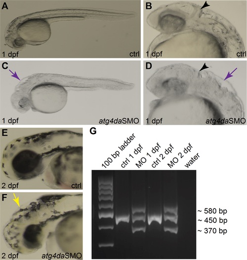
(A-F) Lateral views of control and morphant zebrafish embryos. (A,B) Control embryos appear normal at 1 dpf. (C,D) Morphant embryos show severe abnormalities in different regions of the brain at 1 dpf. Black arrowheads denote the developing cerebellum. Purple arrows indicate the hindbrain irregularities. (E) Control embryo at 2 dpf. (F) A morphant embryo at 2 dpf displaying hydrocephalus (yellow arrow) and small head and eye. (G) RT-PCR assay showing the efficiency of the atg4daSMO. |
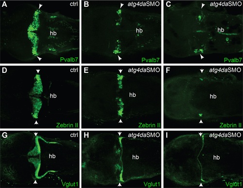
Immunostaining with (A-C) anti-Pvalb7 and (D-F) anti-zebrinII antibody show loss of cerebellar Purkinje cells in atg4da morphants. (A,D) Control embryos at 4.5 dpf. (B,E) Morphants with mild phenotype show partial loss of cerebellar Purkinje cells. (C,F) Morphants with strong phenotype show either total loss of Purkinje cells or presence of few differentiated neurons, which are laterally located in the cerebellum. (G-I) Labeling of cerebellar granule cells with anti-Vglut1 antibody in control and morphant embryos. (H) Mildly affected morphants show reduced expression of Vglut1 in the cerebellum. (I) Vglut1 expression is strongly reduced in embryos showing severe phenotype. White arrowheads indicate the region of cerebellum. Abbreviations: hb, hindbrain. |