
IMAGE
Fig. 5
- ID
- ZDB-IMAGE-150527-5
- Publication
- Kyöstilä et al., 2015 - A Missense Change in the ATG4D Gene Links Aberrant Autophagy to a Neurodegenerative Vacuolar Storage Disease
- All Figures
- Figures for Kyöstilä et al., 2015
Image
|
Figure Caption
Fig. 5
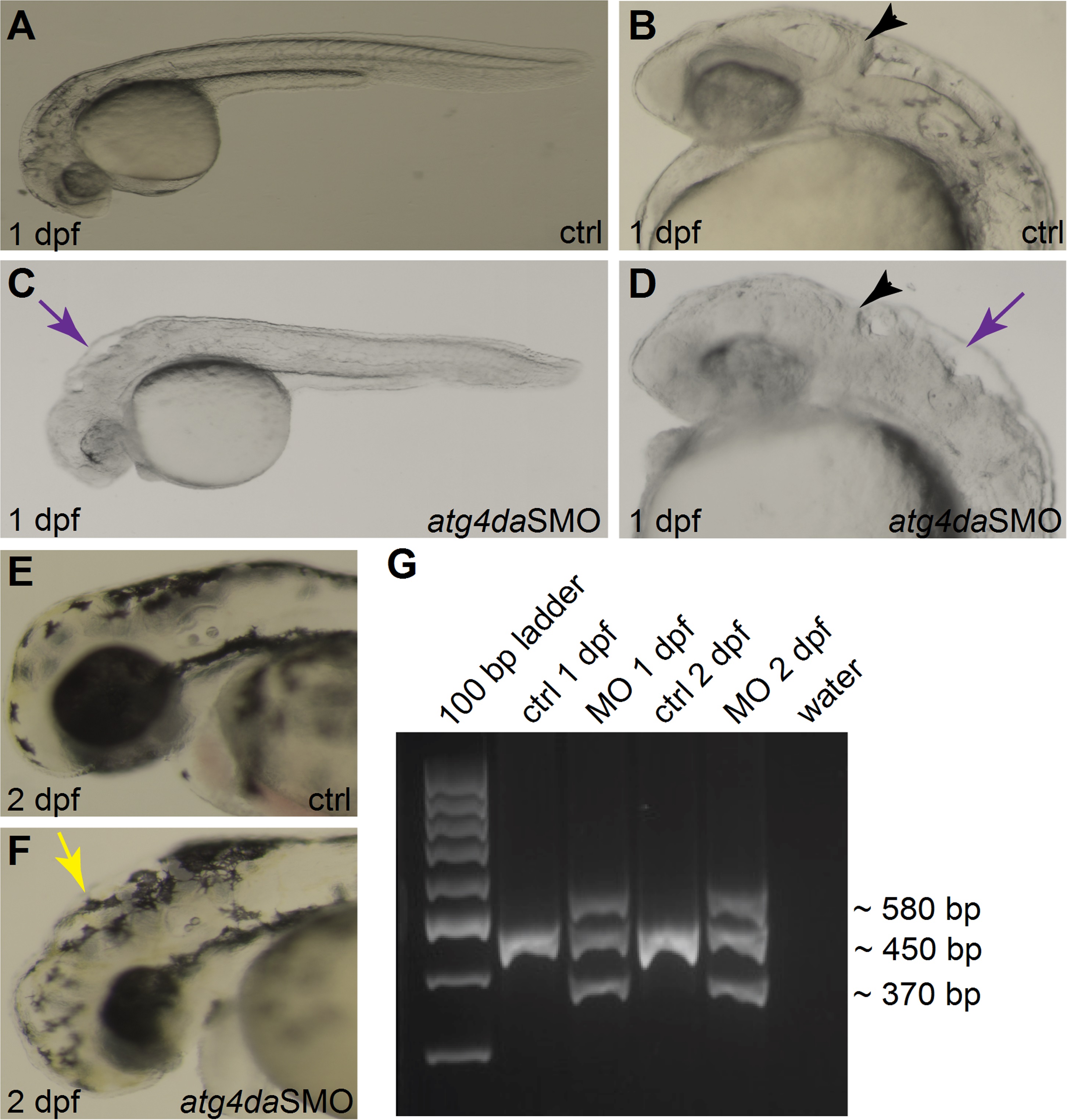
(A-F) Lateral views of control and morphant zebrafish embryos. (A,B) Control embryos appear normal at 1 dpf. (C,D) Morphant embryos show severe abnormalities in different regions of the brain at 1 dpf. Black arrowheads denote the developing cerebellum. Purple arrows indicate the hindbrain irregularities. (E) Control embryo at 2 dpf. (F) A morphant embryo at 2 dpf displaying hydrocephalus (yellow arrow) and small head and eye. (G) RT-PCR assay showing the efficiency of the atg4daSMO.
Figure Data
Acknowledgments
This image is the copyrighted work of the attributed author or publisher, and
ZFIN has permission only to display this image to its users.
Additional permissions should be obtained from the applicable author or publisher of the image.
Full text @ PLoS Genet.