FIGURE SUMMARY
Title

Zebrafish models of cerebrovascular disease

Authors
Walcott, B.P., and Peterson, R.T.
Source
Full text @ J. Cereb. Blood Flow Metab.

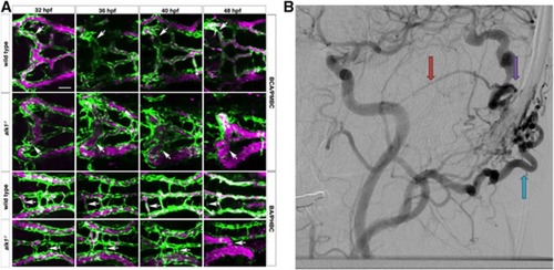
Phenotype comparison of zebrafish and human arteriovenous malformations (AVM). (A) In wild-type embryos (row 1), transient connections between the basal communicating artery (BCA) and primordial midbrain channel (PMBC) carry blood at 32 hpf but regress by 48 hpf (white arrows). In alk1 mutants (row two), one or both of these bilateral connections may be retained, forming an abnormal BCA–to–PMBC arteriovenous connection (white arrows). More posteriorly, lumenized connections drain the basilar artery (BA) to the primordial hindbrain channel (PHBC) in wild-type embryos at early times, but almost all regress by 48 hpf (row 3, white arrows). In alk1 mutants, one or more of these connections may be retained, forming a BA–to–PHBC AVM (row 4, arrows). This model resembles the human condition, seen in a digital subtraction cerebral angiogram. (B) In human AVMs, arterial branches (red arrows) connect directly to the venous circulation (blue arrows) through a high-flow fistula (purple arrow). One theory for AVM development is that they represent the abnormal persistence of normal transient developmental connections. Scale bars, 50 μm. Zebrafish images are two-dimensional confocal projections of Tg(kdrl:GFP)la116; Tg(gata1:dsRed)sd2 embryos, dorsal views, anterior leftwards. Endothelial cells are green; erythrocytes are magenta. Human digital subtraction angiogram is a lateral projection carotid artery injection in the late arterial phase. (Figure and legend modified from Corti P et al.44 Distributed under the terms of the Creative Commons Attribution (CC-BY) License).

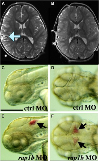
Phenotype comparison of zebrafish and cerebral cavernous malformation. In an magnetic resonance imaging of a human (A), a cerebral cavernous malformation is apparent in the right frontal lobe (blue arrow). These lesions can result in catastrophic hemorrhage and/or seizure activity. Treatment with surgery is effective, and complete resection can be achieved (B) if lesions are in accessible areas. When lesions are deep or multiple, surgical treatment may not be indicated, underscoring the need to develop novel therapeutics. To understand the molecular mechanisms in the CCM pathway, zebrafish models of the disease have been generated using morpholino technology. Compared with control organisms (C and D), morphant knockdowns of rap1b (E and F), a gene that encodes a Ras GTPase effector protein for CCM1/Krit1, demonstrate disrupted endothelial junctions, resulting in intracranial hemorrhage (black arrows), similar to human lesions. Bars, 250 μM. (Figure modified from Gore A V et al.95 Distributed under the terms of the Creative Commons Attribution (CC-BY) License).

Acknowledgments
This image is the copyrighted work of the attributed author or publisher, and ZFIN has permission only to display this image to its users. Additional permissions should be obtained from the applicable author or publisher of the image. Full text @ J. Cereb. Blood Flow Metab.