IMAGE

Figure 2

ID
ZDB-IMAGE-190723-1664
Source
Figures for Walcott et al., 2014
Image
Figure Caption

Figure 2

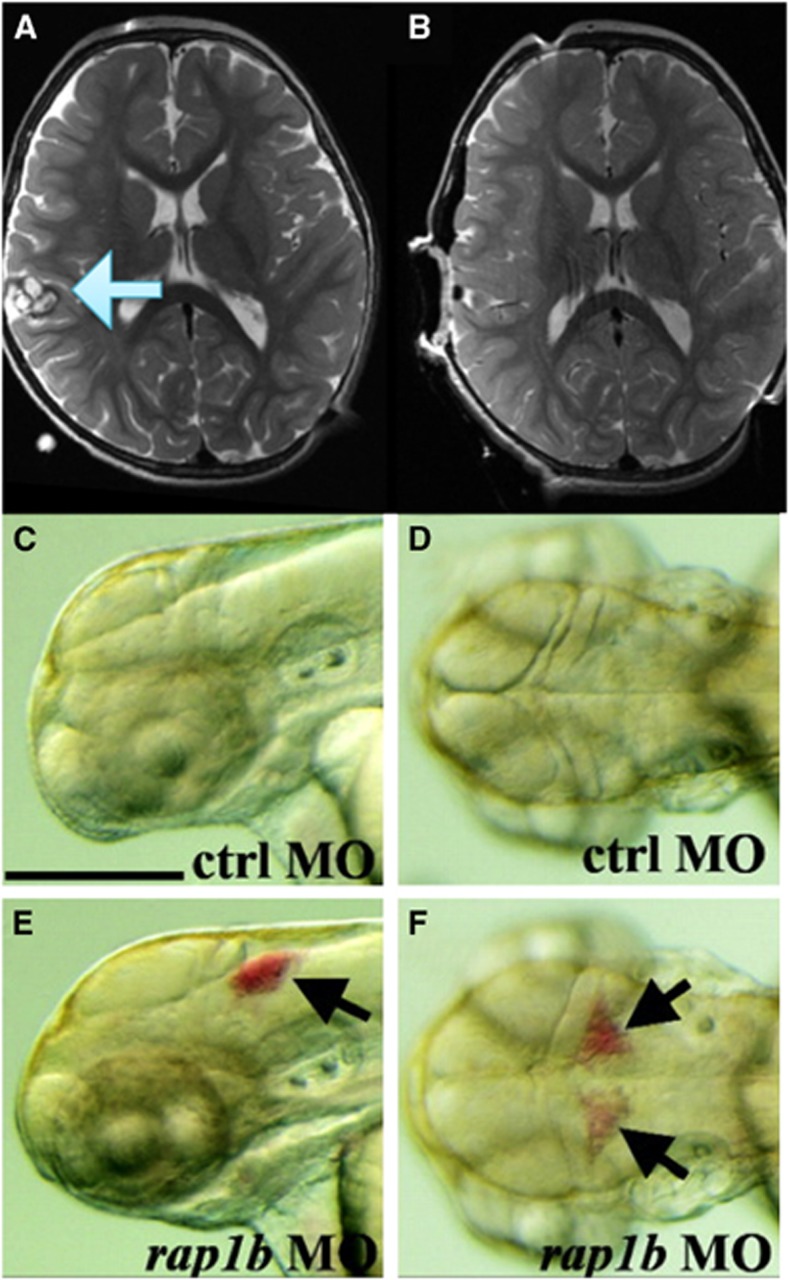
Phenotype comparison of zebrafish and cerebral cavernous malformation. In an magnetic resonance imaging of a human (A), a cerebral cavernous malformation is apparent in the right frontal lobe (blue arrow). These lesions can result in catastrophic hemorrhage and/or seizure activity. Treatment with surgery is effective, and complete resection can be achieved (B) if lesions are in accessible areas. When lesions are deep or multiple, surgical treatment may not be indicated, underscoring the need to develop novel therapeutics. To understand the molecular mechanisms in the CCM pathway, zebrafish models of the disease have been generated using morpholino technology. Compared with control organisms (C and D), morphant knockdowns of rap1b (E and F), a gene that encodes a Ras GTPase effector protein for CCM1/Krit1, demonstrate disrupted endothelial junctions, resulting in intracranial hemorrhage (black arrows), similar to human lesions. Bars, 250 μM. (Figure modified from Gore A V et al.95 Distributed under the terms of the Creative Commons Attribution (CC-BY) License).

Acknowledgments
This image is the copyrighted work of the attributed author or publisher, and ZFIN has permission only to display this image to its users. Additional permissions should be obtained from the applicable author or publisher of the image. Full text @ J. Cereb. Blood Flow Metab.