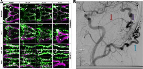
Phenotype comparison of zebrafish and human arteriovenous malformations (AVM). (A) In wild-type embryos (row 1), transient connections between the basal communicating artery (BCA) and primordial midbrain channel (PMBC) carry blood at 32 hpf but regress by 48 hpf (white arrows). In alk1 mutants (row two), one or both of these bilateral connections may be retained, forming an abnormal BCA–to–PMBC arteriovenous connection (white arrows). More posteriorly, lumenized connections drain the basilar artery (BA) to the primordial hindbrain channel (PHBC) in wild-type embryos at early times, but almost all regress by 48 hpf (row 3, white arrows). In alk1 mutants, one or more of these connections may be retained, forming a BA–to–PHBC AVM (row 4, arrows). This model resembles the human condition, seen in a digital subtraction cerebral angiogram. (B) In human AVMs, arterial branches (red arrows) connect directly to the venous circulation (blue arrows) through a high-flow fistula (purple arrow). One theory for AVM development is that they represent the abnormal persistence of normal transient developmental connections. Scale bars, 50 μm. Zebrafish images are two-dimensional confocal projections of Tg(kdrl:GFP)la116; Tg(gata1:dsRed)sd2 embryos, dorsal views, anterior leftwards. Endothelial cells are green; erythrocytes are magenta. Human digital subtraction angiogram is a lateral projection carotid artery injection in the late arterial phase. (Figure and legend modified from Corti P et al.44 Distributed under the terms of the Creative Commons Attribution (CC-BY) License).
|
