
- Title
-
Conserved roles of the prion protein domains on subcellular localization and cell-cell adhesion
- Authors
- Solis, G.P., Radon, Y., Sempou, E., Jechow, K., Stuermer, C.A., and Málaga-Trillo, E.
- Source
- Full text @ PLoS One

Accumulation of mouse and zebrafish PrPs at newly formed cell contacts in Drosophila S2 cells. A) Expression of the mouse PrP EGFP fusion construct (m PrP) induces cell contact formation and the subsequent accumulation of PrP at contact sites. This is not observed at fortuitous contacts formed by cells expressing a control construct lacking the major PrP domains (m PrP ΔCore). Cell-cell contacts are indicated by white arrowheads. Scale bars = 5 μm. B-D) Quantification of the number of S2 cell contacts showing accumulation of wild type (WT) and mutant constructs for mouse PrP (B), zebrafish PrP-1 (C) and zebrafish PrP-2 (D). Construct names are inserted in the graphs. Double and triple asterisks [** and ***] indicate statistical significance at p<0.01 and <0.001, respectively; one-way ANOVA test; error bars indicate SEM. |
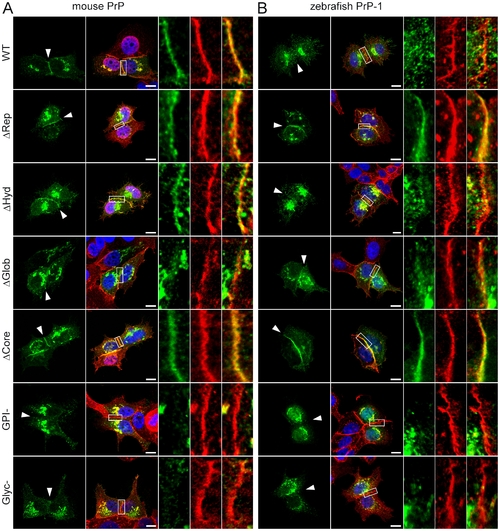
Accumulation of mouse and zebrafish PrP constructs at established MCF-7 cell cell contacts. Wild type (WT) and mutant EGFP-tagged constructs of mouse PrP (A) and zebrafish PrP-1 (B) localize differently at E-cadherin-positive cell contact sites (in red). Marked areas on the overlays are enlarged (right) to show detailed views of the contact sites. Cell nuclei are stained with DAPI (blue). Scale bars = 10 μm. |
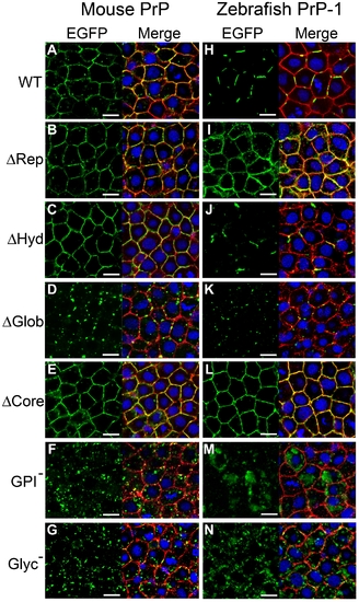
Localization of mouse PrP and zebrafish PrP-1 constructs in early zebrafish embryos. Expression of EGFP-tagged constructs (green) in the deep cells of 6 hpf zebrafish gastrulae. Plasma membranes were double-counterstained using antibodies against pY416-Src and β-catenin (merged in red). Cell nuclei were stained with DAPI (blue). Scale bars = 10 μm. |
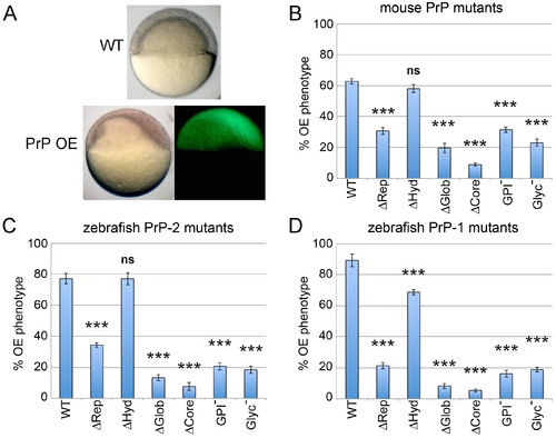
Overexpression (OE) of mouse and zebrafish PrP constructs in early zebrafish embryos. Embryos were microinjected with mRNAs encoding all mouse and zebrafish EGFP-tagged PrP constructs. A) OE of mouse or zebrafish WT PrPs in early embryos produces a gain-of-function phenotype characterized by asymmetric gastrulation at 6 hpf. B–D) The activities of mouse and zebrafish constructs were evaluated morphologically by quantifying the proportion of 6 hpf embryos exhibiting the OE phenotype (asymmetric gastrulation). Three independent experiments were analyzed (average n = 30 embryos). Triple asterisks [***] indicate statistically significant reduction in activity at p<0.001; one-way ANOVA test; error bars represent SEM. |
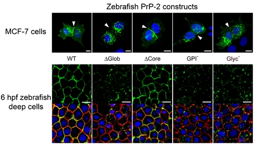
Localization of zebrafish PrP-2 at cell-cell contacts in epithelial MCF-7 cells and 6 hpf zebrafish deep cells. Expression of zebrafish PrP-2 EGFP fusion wild type (WT) and mutant (indicated in the figures) constructs localized differentially at cell contact sites (white arrowheads). Scale bars = 10 μm. |