
Image
|
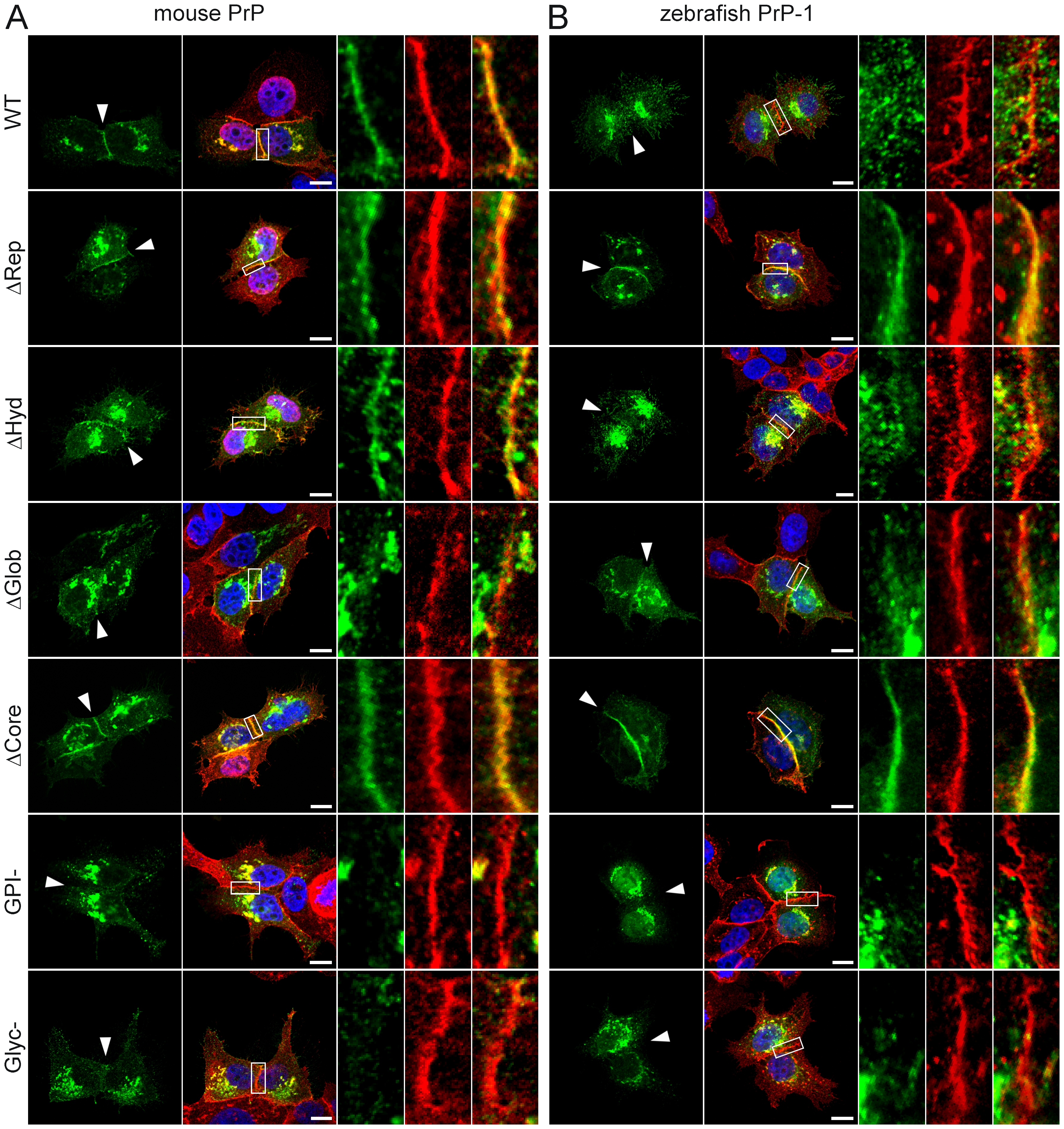
Figure Caption
Fig. 3
Accumulation of mouse and zebrafish PrP constructs at established MCF-7 cell cell contacts.
Wild type (WT) and mutant EGFP-tagged constructs of mouse PrP (A) and zebrafish PrP-1 (B) localize differently at E-cadherin-positive cell contact sites (in red). Marked areas on the overlays are enlarged (right) to show detailed views of the contact sites. Cell nuclei are stained with DAPI (blue). Scale bars = 10 μm.
Acknowledgments
This image is the copyrighted work of the attributed author or publisher, and
ZFIN has permission only to display this image to its users.
Additional permissions should be obtained from the applicable author or publisher of the image.
Full text @ PLoS One