FIGURE SUMMARY
Title

Engineering the Biosynthesis of the Polyketide-Nonribosomal Peptide Collismycin A for Generation of Analogs with Neuroprotective Activity

Authors
Garcia, I., Vior, N.M., González-Sabín, J., Braña, A.F., Rohr, J., Moris, F., Méndez, C., and Salas, J.A.
Source
Full text @ Chem. Biol.

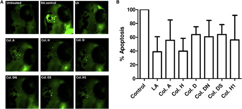
Neuroprotective Action of the Collismycin Analogs
(A) Induction of apoptosis in a zebrafish model as visualized by fluorescence microscopy. The zebrafish larvae were treated with 10 μM RA alone as a stress inducer (RA control) or in combination with 1 μM of the different collismycin analogs. As a positive neuroprotectant control, zebrafish were treated with 10 μM RA and 1 μM LA. See also Table S2.
(B) Effect of the different collismycin analogs on RA-induced apoptosis in zebrafish larvae. Apoptosis induced by RA in the presence or absence of different collismycin analogs was determined with fluorescence microscopy and, after quantification, relative apoptosis was standardized using the level of apoptosis generated by the RA treatment as 100%. One-way ANOVA and Dunnett’s t test were used to determine the significance. A p value less than 0.05 was considered statistically significant. All analogs exhibited significant protection with p < 0.05. See also Table S1.
See also Figures S27–S29.

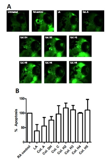
"Figure S29, related to Figure 5". Zebrafish assay results for compounds Collismycin DH, Collismycin C, Collismycin H2, Collismycin H3, Collismycin H4 and Collismycin H5.
(A): Induction of apoptosis in the zebrafish model visualized by fluorescence microscopy. The zebrafish larvae were treated with 10 μM retinoic acid alone as stress inducer (RA control) or in combination with 1 μM of the different collismycin analogs. As a positive neuroprotectant control zebrafish were treated with 10 μM RA and 1 μM lipoic acid (LA).
(B): Effect of the different collismycin analogs on retinoic acid-induced apoptosis in zebrafish larvae. Apoptosis induced by retinoic acid in the presence or absence of collismycin A and different collismycin analogs was determined by fluorescence microscopy and, after quantification, relative apoptosis was standardized using the level of apoptosis generated by the RA treatment as 100%. None of the analogs here represented exhibited significant protection.

Acknowledgments
This image is the copyrighted work of the attributed author or publisher, and ZFIN has permission only to display this image to its users. Additional permissions should be obtained from the applicable author or publisher of the image. Full text @ Chem. Biol.