
- Title
-
Aminoglycoside-Induced Hair Cell Death of Inner Ear Organs Causes Functional Deficits in Adult Zebrafish (Danio rerio)
- Authors
- Uribe, P.M., Sun, H., Wang, K., Asuncion, J.D., Wang, Q., Chen, C.W., Steyger, P.S., Smith, M.E., and Matsui, J.I.
- Source
- Full text @ PLoS One
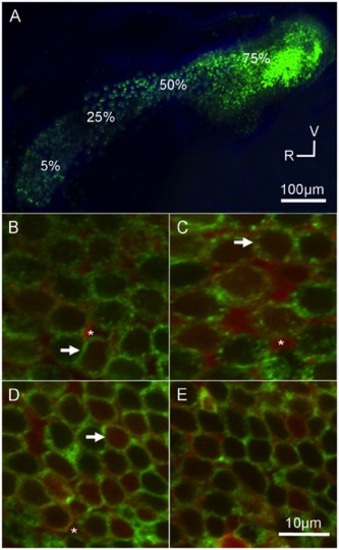
Gentamicin localizes to saccular hair cells. Adult Brn3c-GFP zebrafish received an intraperitoneal injection of Texas Red-conjugated gentamicin (GTTR; 30 mg/kg) and allowed to recover for 4 hours. (A) Low magnification image of the saccule. GFP-labeled hair cells (green) are located along the length of the organ. V = ventral, R = rostral. High magnification images (B–E) are single optical slices from individual z-stacks taken at (B) 5%, (C) 25%, (D) 50%, and (E) 75% along the length of the saccule from the rostral end (see A). GTTR fluorescence (red) was localized to hair cells (D, white arrows) and supporting cells (white asterisks) throughout the sensory epithelium. GFP fluorescence (green) is localized to the cell membrane region of hair cells. The z-stack slices are taken mid-way into the cell to show the localization patterns, thus neither hair cell nuclei nor stereociliary bundles are visible. |

Gentamicin localizes to utricular hair cells. Adult Brn3c-GFP zebrafish received an intraperitoneal injection of GTTR (30 mg/kg) and allowed to recover for 4 hours. (A) Low magnification image of the utricle. GFP-labeled hair cells (green) are located throughout the organ. The striolar region (S) and extrastriolar region (E) are highlighted. High magnification images (B, C) are single optical slices from individual z-stacks from (B) striolar and (C) extrastriolar regions. GTTR fluorescence (red) was localized to hair cells (white arrows) and supporting cells (white asterisks) throughout the sensory epitheilum. GFP fluorescence (green) is localized to the cell membrane region of hair cells. The slices are taken mid-way through the cell to show the localization patterns, thus, neither hair cell nuclei nor stereociliary bundles are visible. |

ZFIN is incorporating published figure images and captions as part of an ongoing project. Figures from some publications have not yet been curated, or are not available for display because of copyright restrictions. PHENOTYPE:
|
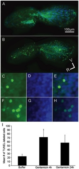
Gentamicin treatment increases the numbers of TUNEL-labeled cells in the adult saccule. Adult wildtype zebrafish received either a single intraperitoneal injection of (A, C, D, E) buffer or (B, F, G, H) 250 mg/kg gentamicin and allowed to recover for either 4 or 24 hours (4 hour data is shown). Fixed saccules were processed using a TUNEL-kit to label apoptotic cells (green cells; A, B, C, E, F, H) and co-labeled with DAPI (blue cells; A, B, D, E, G, H). (A-B) Low power and (C–H) high power images of the saccule reveals few TUNEL-labeled cells scattered throughout the saccule of a buffer-treated animal (A, C, E) but more TUNEL-labeling in a gentamicin-treated animal (B, F, H). (I) Mean number of TUNEL-labeled cells (± SD) in the saccules of buffer- or gentamicin-injected zebrafish. *p<0.05; n = 9–12; V = ventral, R = rostral. PHENOTYPE:
|
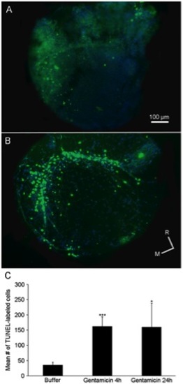
Gentamicin treatment increases the numbers of TUNEL-labeled cells in the adult utricle. Adult wildtype zebrafish received either a single intraperitoneal injection of (A) buffer or (B) 250 mg/kg gentamicin and allowed to recover for either 4 or 24 hours (4 hour data is shown). Fixed utricles were processed using a TUNEL-kit to label apoptotic cells (green cells) and co-labeled with DAPI (blue cells). (A–B) Low power (10X) images of the utricle reveals few TUNEL-labeled cells scattered throughout the utricle of a (A) buffer-treated animal but more cells with TUNEL-labeling in the (B) gentamicin-treated animal. (C) Mean number of TUNEL-labeled cells (± SD) in the utricles of buffer- or gentamicin-injected zebrafish. *p<0.05 or ***p<0.001; n = 9–12; M = medial, R = rostral. PHENOTYPE:
|
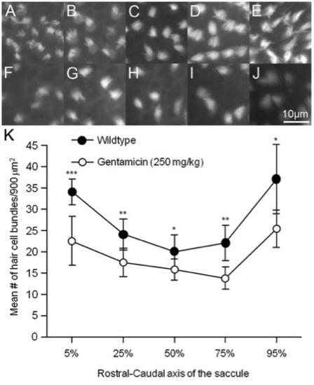
Gentamicin treatment resulted in fewer saccular hair bundles after 24 hours. Adult wildtype zebrafish received a single 250 mg/kg intraperitoneal injection of gentamicin and allowed to recover for 24 hours. Images of phalloidin-labeled cells were taken at (A, F) 5%, (B, G) 25%, (C, H) 50%, (D, I) 75%, and (E, J) 95% along the length of the saccule beginning at the rostral end in (A–E) untreated controls and (F–J) gentamicin-treated animals. (K) Mean (± SD) number of saccular hair cell bundles per 900 μm2 of epithelia. Significantly fewer phalloidin-labeled hair bundles were counted at each area along the length of the saccule in gentamicin-treated animals compared to controls. *p<0.05 or **p<0.01; n = 6–13 saccules per area per condition. PHENOTYPE:
|
|
Unillustrated author statements PHENOTYPE:
|