
- Title
-
Expression of the AMACO (VWA2 protein) ortholog in zebrafish
- Authors
- Gebauer, J.M., Karlsen, K.R., Neiss, W.F., Paulsson, M., and Wagener, R.
- Source
- Full text @ Gene Expr. Patterns
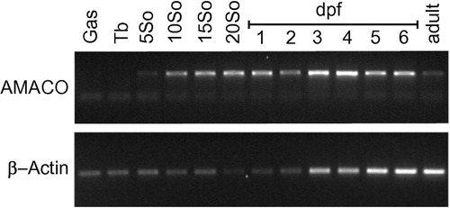
RT-PCR analysis of AMACO mRNA species expressed during zebrafish development. RT-PCR analysis was performed using the primer pair zAMACOp3 fw and rev. β-Actin was used as a loading control. Gas, gastrulation; Tb, tail bud; So, somite; dpf, days past fertilization. EXPRESSION / LABELING:
|
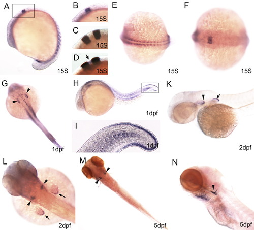
The expression pattern of AMACO transcripts during the development of zebrafish larvae. Zebrafish embryos at the 15 somite-stage (15S) (A–F), at 1dpf (G–I), 2dpf (K,L) and 5dpf (M,N) were hybridized with probes specific for AMACO (A, B, D–N) or krox-20 (C,D). All pictures were taken with a Nikon stereomicroscope, except for (I), which was taken with a Zeiss Axiophot. At the 15 somite stage the AMACO gene is transcribed in the somites (A,E) and in the hindbrain (A, B, F). (A–D) Lateral view. (E) Dorsal view of the somites and (F) dorsal view of the hindbrain, anterior to the left. The box in (A) delineates the area that is enlarged in (B–D). To assess the exact localization of AMACO transcription in the hindbrain at the 15 somite stage (A, B, F), a double in situ hybridization with riboprobes for AMACO and krox-20, which labels rhombomeres 3 and 5 (Sun et al., 2002), was performed. Although much weaker, a clear AMACO signal (arrow) that is lacking in the single krox-20 staining (C) is seen in the double staining (D). At 1dpf AMACO is expressed in the otic vesicle (arrowheads), the somites, the tail fin fold and at the lining of brain ventricles (G–I). The box in H delineates the area that is shown enlarged in (I), which shows that the somite borders are not stained. (G) Dorsal view, (H,I) lateral views. At 2 dpf AMACO is expressed in the otic capsule (arrowheads) and the pectoral fin (arrows), (K) lateral view, (L) dorsal view. At 5 dpf AMACO is also expressed in the gills, in addition to in the otic capsule (arrowheads), (M) dorsal view, (N) lateral view. EXPRESSION / LABELING:
|
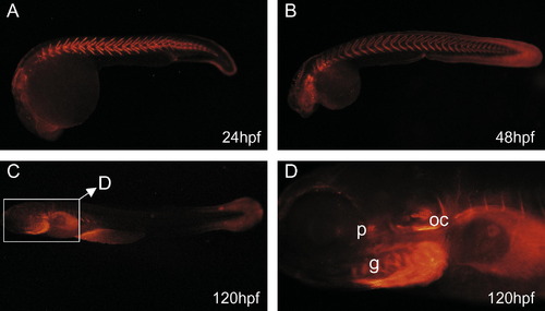
AMACO distribution in zebrafish larvae. Immunofluoresence microscopy was carried out after whole-mount antibody staining. Zebrafish embryos were incubated with the affinity-purified antibody directed against AMACO followed by Alexa 546-conjugated goat anti-rabbit IgG. At 24 hpf AMACO is mainly expressed between the somites (A). At later stages (B,C) deposition between the somites decreases and additional signals can be detected at the fin fold (>48 hpf, (B)), the gills (g), the otic capsule (oc) and the pituitary gland (p) (D). All pictures show lateral views, anterior to the left. |
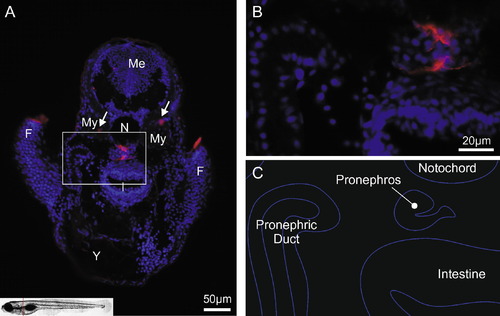
AMACO expression in the pronephros. Immunofluoresence microscopy was carried out on paraffin-embedded tissue sections from 4-day-old zebrafish using an affinity-purified antibody directed against zebrafish AMACO followed by Alexa 546-conjugated goat anti-rabbit IgG. The red line in the picture of a zebrafish in the lower left corner of A indicates the position of the lateral section that is shown in (A,B). (A) AMACO is mainly expressed in basement membrane structures of the pronephros. Signals were also observed in the fin folds. Arrows indicate expression in myosepta. AMACO: red; nuclear staining: blue; (B) is a magnification of boxed region in (A): (C) is a schematic drawing of organs seen in (B). F, fin fold; Me, mesencephalon; My myotome; N, notochord; I, intestine; Y, yolk. EXPRESSION / LABELING:
|
Reprinted from Gene expression patterns : GEP, 10(1), Gebauer, J.M., Karlsen, K.R., Neiss, W.F., Paulsson, M., and Wagener, R., Expression of the AMACO (VWA2 protein) ortholog in zebrafish, 53-59, Copyright (2010) with permission from Elsevier. Full text @ Gene Expr. Patterns