
IMAGE
Fig. 5
- ID
- ZDB-IMAGE-100302-32
- Genes
- Antibodies
- Publication
- Gebauer et al., 2010 - Expression of the AMACO (VWA2 protein) ortholog in zebrafish
- All Figures
- Figures for Gebauer et al., 2010
Image
|
Figure Caption
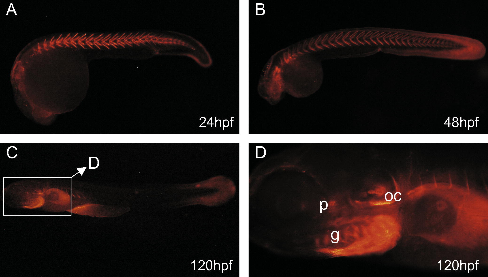
Fig. 5 AMACO distribution in zebrafish larvae. Immunofluoresence microscopy was carried out after whole-mount antibody staining. Zebrafish embryos were incubated with the affinity-purified antibody directed against AMACO followed by Alexa 546-conjugated goat anti-rabbit IgG. At 24 hpf AMACO is mainly expressed between the somites (A). At later stages (B,C) deposition between the somites decreases and additional signals can be detected at the fin fold (>48 hpf, (B)), the gills (g), the otic capsule (oc) and the pituitary gland (p) (D). All pictures show lateral views, anterior to the left.
Figure Data
Acknowledgments
This image is the copyrighted work of the attributed author or publisher, and
ZFIN has permission only to display this image to its users.
Additional permissions should be obtained from the applicable author or publisher of the image.
Reprinted from Gene expression patterns : GEP, 10(1), Gebauer, J.M., Karlsen, K.R., Neiss, W.F., Paulsson, M., and Wagener, R., Expression of the AMACO (VWA2 protein) ortholog in zebrafish, 53-59, Copyright (2010) with permission from Elsevier. Full text @ Gene Expr. Patterns