
Fig. 6
- ID
- ZDB-IMAGE-100302-33
- Genes
- Antibodies
- Publication
- Gebauer et al., 2010 - Expression of the AMACO (VWA2 protein) ortholog in zebrafish
- All Figures
- Figures for Gebauer et al., 2010
|
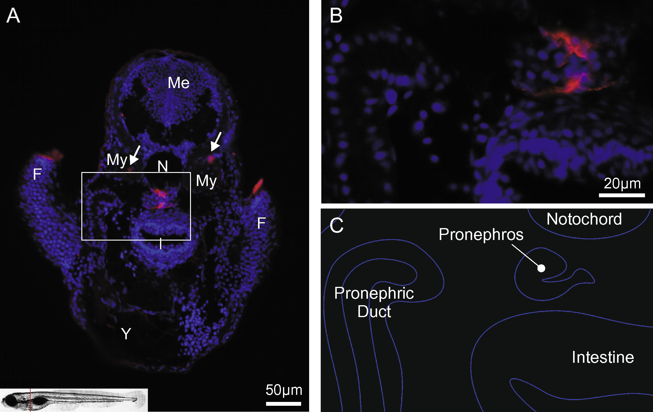
Fig. 6 AMACO expression in the pronephros. Immunofluoresence microscopy was carried out on paraffin-embedded tissue sections from 4-day-old zebrafish using an affinity-purified antibody directed against zebrafish AMACO followed by Alexa 546-conjugated goat anti-rabbit IgG. The red line in the picture of a zebrafish in the lower left corner of A indicates the position of the lateral section that is shown in (A,B). (A) AMACO is mainly expressed in basement membrane structures of the pronephros. Signals were also observed in the fin folds. Arrows indicate expression in myosepta. AMACO: red; nuclear staining: blue; (B) is a magnification of boxed region in (A): (C) is a schematic drawing of organs seen in (B). F, fin fold; Me, mesencephalon; My myotome; N, notochord; I, intestine; Y, yolk.
Reprinted from Gene expression patterns : GEP, 10(1), Gebauer, J.M., Karlsen, K.R., Neiss, W.F., Paulsson, M., and Wagener, R., Expression of the AMACO (VWA2 protein) ortholog in zebrafish, 53-59, Copyright (2010) with permission from Elsevier. Full text @ Gene Expr. Patterns