
- Title
-
A novel puf-A gene predicted from evolutionary analysis is involved in the development of eyes and primordial germ-cells
- Authors
- Kuo, M.W., Wang, S.H., Chang, J.C., Chang, C.H., Huang, L.J., Lin, H.H., Yu, A.L., Li, W.H., and Yu, J.
- Source
- Full text @ PLoS One
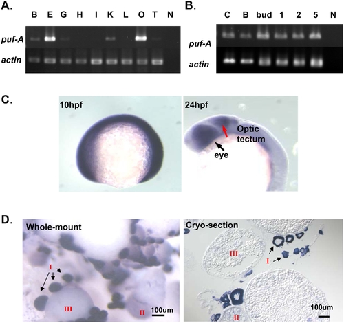
Expression of puf-A in zebrafish using RT-PCR and in situ hybridization. (A) Gene expression in adult tissues of zebrafish was analyzed by RT-PCR and electrophoresis with puf-A primers (upper panel) or actin primers (lower panel, as the internal control). Notation: B, brain; E, eye; G, gill; H, heart; I, intestine; K, head kidney; L, liver; O, ovary; T, testis; N, negative control. (B) The puf-A gene was expressed in various stages of zebrafish embryo. C, cleavage; B, blastula; 1, 1 day post-fertilization (dpf); 2, 2 dpf; 5, 5 dpf; N, negative control. (C) Whole-mount in situ hybridization with puf-A antisense riboprobe on zebrafish embryo. The puf-A expression at 10 h post-fertilization (hpf) (tailbud stage) and 24 hpf (25-somite stage) with lateral overview. The black arrow points to the eye and the red arrow to the optic tectum. (D) Whole-mount and cryo-section in situ hybridization with a puf-A antisense riboprobe in adult ovaries. In adult ovaries, a staging series of oocyte development was characterized by the diameter of various oocytes [9], [10]. Stage I, primary growth follicles (<0.1 mm); stage II, previtellogenic (0.1–0.30 mm); and stage III, vitellogenic (>0.30 mm). |
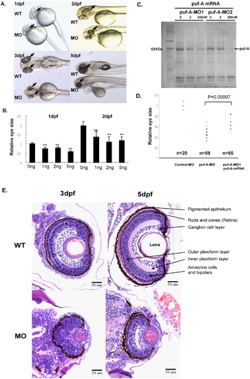
The phenotypes of puf-A morphants in the zebrafish. (A) Zebrafish embryos at the 1∼4-cell stage were treated with 5 ng puf-A morpholino (MO1) by microinjection. The phenotypes of the wild-type and morphants are shown in lateral view at 1, 2, 3, and 5 days post-fertilization (dpf) after treatment. Black arrows point to the eyes. (B) Various amounts of MO1 were microinjected into zebrafish embryos, and the eye size was measured at 1 or 2 dpf and compared to the eye size of control fish. The “relative eye size” was defined by the value of eye size in MOs relative to the average size of eyes in normal embryos of WT fish. The average value of eye size in normal embryos at 1dpf was considered as 1. Error bars represent the standard error of the mean. ** refer to p<0.01 Student's t-test). (C) 0, 2, 200 nM puf-A-MO1 or puf-A-MO2 were added to the puf-A generated through in vitro transcription/translation reactions. One microliter of the reaction mixture was separated on 10% SDS/PAGE, blotted, incubated with streptavidin-AP, and developed with NBT-BCIP reagents. (D) The 5 ng control- or puf-A-MO1 was used for microinjection. In addition, 200 pg of capped puf-A RNA was co-injected with puf-A-MO1 to check the specificity of MO knockdown. The “relative eye size” was defined as above. The eye size was measured at 2 dpf. Control-MO, n = 20 embryos; puf-A-MO1, n = 59; puf-A-MO1+ capped RNA, n = 65. The p value in Student's t-test for the difference between puf-A-MO and puf-A-MO+mRNA was <0.00007. (E) Transverse histological sections of zebrafish wild-type and morphant (puf-A-MO1) eyes stained with hematoxylin and eosin at 3 and 5 dpf. |
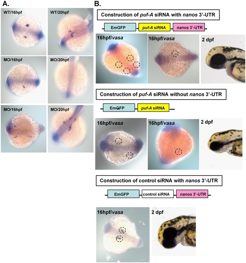
The puf-A gene plays a role in the PGC development in zebrafish embryonic development. (A) The vasa antisense riboprobe was used in whole-mount in situ hybridization as a marker to monitor primordial germ-cell (PGC) development in zebrafish. The vasa expression in wild-type (WT) and morphants (MO1 at 5 ng/embryo) at 16 and 20 hpf embryos in dorsal view. Anterior is to the left for 16 hpf and left bottom for 20 hpf. The morphants exhibited prominent abnormalities with either a reduction in PGC numbers (50.4%, n = 125 embryos) or abnormal patterns of migration (34.4%, n = 125) indicating the failure of PGC navigation towards their destined sites. (B) Upper: the construction of puf-A siRNA with nanos 3′ UTR, vasa expression and normal eye size in embryos 2 dpf after injection. The embryos displayed a marked reduction in PGC numbers (80.9%, n = 115 embryos) and abnormal patterns of migration (11.3%, n = 115 embryos). Middle: construction of puf-A siRNA without nanos 3′ UTR, vasa expression and small eye size in 2dpf embryos. Bottom: construction of control siRNA with nanos 3′ UTR, vasa expression and normal eye size in embryos after injection. EXPRESSION / LABELING:
PHENOTYPE:
|
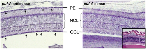
The puf-A gene was expressed in adult murine eyes. Panels show in situ hybridization of an adult mouse eye with NBT-BCIP color reaction after hybridization with puf-A antisense and sense probes, separately. The black arrows point to puf-A expression which was probed with antisense, while mouse sense probe served as the negative control. HE staining inside the mouse sense panel shows the different retinal layers of the mouse eye. PE, pigmented epithelium; NCL, nuclear cell layer. |
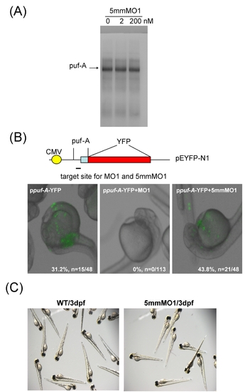
The in vitro and in vivo analyses for the specificity of puf-A MO1. (A) Various amounts of puf-A-5mmMO1 (5 bp mismatch control: 0, 2, and 200 nM) were added to the in vitro transcription/translation reactions for puf-A. One microliter of the reaction mixture was separated on 10% SDS/PAGE, blotted and incubated with streptavidin-AP, followed by development with NBT-BCIP reagents.(B) For in vivo experiment, the puf-A 5′-UTR and its partial coding region were added onto pEYFP-N1 plasmid which contained CMV promoter and YFP gene to generate ppuf-A-YFP. Then 4.6 ng of the puf-A-MO1 or puf-A-5mmMO1 was co-injected to embryo with ppuf-A-YFP plasmid at 100 pg per embryo. The numbers of embryo with YFP expression were enumerated at 1dpf. As shown, 15/48 embryos injected puf-A-YFP plasmid exhibited fluorescence at 100 pg/embryo dosage, while none of the 113 embryos co-injected with the puf-A-MO1 had YFP expression. In contrast, co-injection with the mismatch control, puf-A-5mmMO1, did not affect the expression of YFP. (C) The panels showed the phenotypes of WT and morphants at 3dpf after injection with 9.2 ng of puf-A-5mmMO1 dosage. |
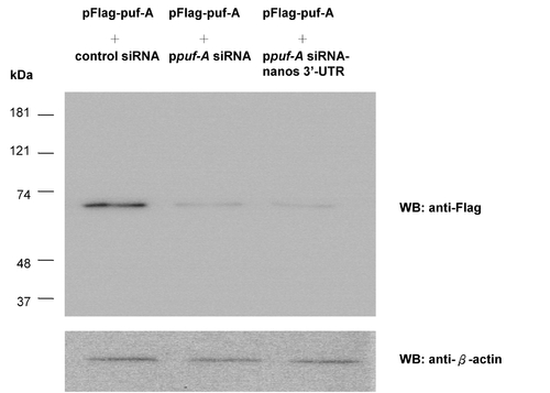
The puf-A siRNA suppressed specifically the zebrafish puf-A expression in 3T3 cell line. The 3T3 cell line was co-transfected with pFlag-puf-A and different siRNAs. In the first line of Western blot, the siRNA was the control siRNA (pcDNA 6.2-GW/EmGFP-miR -neg control plasmid) as negative control. In the middle line, ppuf-A siRNA and in the last line, ppuf-A siRNA containing nanos 3′-UTR were used to suppress the puf-A expression. Upper panel showed the Western blot after reaction with anti-Flag antibodies, while lower panel showed Western blot for β-actin as internal control. |
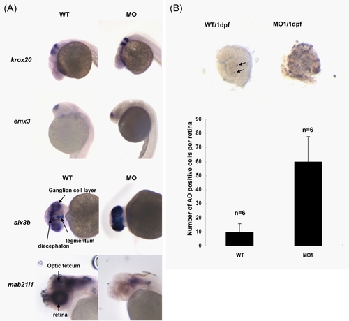
Expression of in situ marker genes and elevated level of apoptosis in puf-A morphants (A) The upper four panels showed normal expression of krox20 and emx3 in WT and morphants, separately, at 1dpf. The lower four panels showed the abnormal expression patterns of six3b and mab21l1 in WT and morphants at 2dpf. (B) Apoptotic cells were detected by terminal deoxynucleotidyl transferase-mediated dUTP nick end labeling (TUNEL) using an In Situ Cell Death Detection kit (Roche). Embryos were fixed with 4% PFA and whole eyes at 1dpf were dissected out. Black arrows refer to the apoptotic cells in eyes of WT embryos. Acridine orange (AO) was also used to label apoptotic cells in zebrafish embryos. The average number of AO positive cells per retina in wild-type (WT, n = 8) and puf-A morphants (MO1, n = 8) at 1dpf was presented. |