
Fig. S5
- ID
- ZDB-FIG-090401-45
- Publication
- Kuo et al., 2009 - A novel puf-A gene predicted from evolutionary analysis is involved in the development of eyes and primordial germ-cells
- Other Figures
- All Figure Page
- Back to All Figure Page
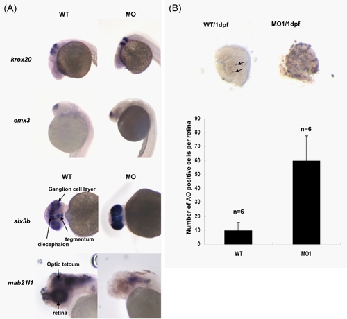
Expression of in situ marker genes and elevated level of apoptosis in puf-A morphants (A) The upper four panels showed normal expression of krox20 and emx3 in WT and morphants, separately, at 1dpf. The lower four panels showed the abnormal expression patterns of six3b and mab21l1 in WT and morphants at 2dpf. (B) Apoptotic cells were detected by terminal deoxynucleotidyl transferase-mediated dUTP nick end labeling (TUNEL) using an In Situ Cell Death Detection kit (Roche). Embryos were fixed with 4% PFA and whole eyes at 1dpf were dissected out. Black arrows refer to the apoptotic cells in eyes of WT embryos. Acridine orange (AO) was also used to label apoptotic cells in zebrafish embryos. The average number of AO positive cells per retina in wild-type (WT, n = 8) and puf-A morphants (MO1, n = 8) at 1dpf was presented. |