FIGURE SUMMARY
Title

Identification of genetic and chemical modulators of zebrafish mechanosensory hair cell death

Authors
Owens, K.N., Santos, F., Roberts, B., Linbo, T., Coffin, A.B., Knisely, A.J., Simon, J.A., Rubel, E.W., and Raible, D.W.
Source
Full text @ PLoS Genet.

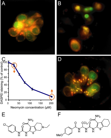
Screening for modifiers of aminoglycoside toxicity.
(A) Neuromast from a control animal pretreated with 0.5% DMSO and stained with rapidly with FM 1-43FX (red) and the nuclear label Yo-Pro-1 (green). (B) Negative control pretreated with 0.05% DMSO for 1 hour followed by 200 μM neomycin treatment for 30 min. Hair cells are stained with FM 1-43FX (red) and Yo-Pro-1 (green). Hair cell loss, nuclear condensation and cytoplasmic shrinking are observed. (C) Dose-response function showing decreased hair cell labeling with DASPEI, a mitochondrial potentiometric dye, as a function of increasing neomycin concentration for wildtype zebrafish (N = 25–37 total fish per group, from triplicate experiments). Bars are SEM. Screens for increased or decreased susceptibility to hair cell loss were performed by treatment with either low, 25 μM, or high, 200 μM, neomycin doses, respectively, as highlighted by the orange arrows. (D) Neuromast pretreated with PROTO-2, a compound identified to provide protection against 200 µM neomycin exposure. (E,F) Show the structure for the identified compounds, PROTO-1 (E) and PROTO-2 (F), respectively.

Mutations that confer protection against neomycin exposure.
Larvae are labeled with DASPEI after 30 min exposure to 200 μM neomycin and 1 hr recovery in normal media. (A) Wildtype animal shows retention of hair cells in neuromasts after mock-treatment. (B) Wildtype animal shows loss of hair cells after aminoglycoside treatment. (C) persephone mutant animal shows robust protection of neuromasts against neomycin treatment. No morphological defects are observed. (D) sentinel mutant animal shows protection, along with sinusoidal body curvature. Bar = 200 μm.

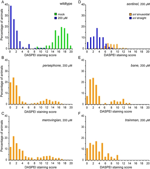
Hair cell retention after neomycin treatment in wildtype and mutant animals.
Histograms show the fraction of animals with different levels of DASPEI staining. For each animal, 10 specific neuromasts are evaluated and assigned a score of 2 (normal staining), 1 (reduced staining), or 0 (no staining) for a maximum total score of 0–20. For each group, the distribution of animals given each DASPEI staining score is displayed as a percentage of the total number of animals to illustrate the phenotypic variation within the group; 40–80 animals were tested for each group. (A) Distribution of wildtype fish after mock treatment without neomycin (green bars) or after exposure to 200 μM neomycin (blue bars). (B–F) Distribution of progeny from crosses between heterozygous mutant carriers treated with 200 μM neomycin, showing both wildtype and mutant phenotypes. (B) persephone. (C) merovingian. (D) sentinel. Animals with sinusoidal bodies (later shown to be homozygous mutants) are represented by orange bars, and animals with wildtype body shape (wildtype or heterozygous siblings) are represented by blue bars. (E) bane. (F) trainman.

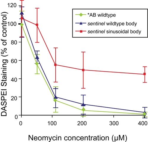
Dose dependent protection of sentinel mutants to neomycin.
Hair cell loss as determined by DASPEI staining of progeny of sentinel heterozygous parents with wildtype body shape (blue) or sinusoidal body shape (red) are compared to wildtype *AB fish (green). Error bars are ±1 S.D. Mutants show robust, but partial, protection following 30 min neomycin exposure and one hour recovery.

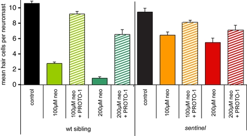
Epistasis analysis of sentinel and protective compounds.
Neomycin dose-response relationship showing effects of 10 μM PROTO-1 against 100 μM or 200 μM neomycin exposure in wildtype and sentinel larvae. For each group, hair cells were pre-labeled with FM1-43FX. Animals were pretreated with PROTO-1 for 1 hour (or mock-treated), treated with neomycin and PROTO-1 for 1 hour, euthanized, and fixed. Hair cells of four neuromasts (left and right) were counted and the average number of hair cells per neuromast was determined. Number of hair cells in control animals (no PROTO-1, no neomycin) are shown with black bars, animals treated with only 100 μM or 200 μM neomycin are shown by solid colored bars, and animals treated with PROTO-1 and neomycin are shown by hatched colored bars. Error bars show 1 S.E.M. PROTO-1 and sentinel mutants show similar protection, and there is a small, statistically significant effect of the combined treatment of the mutation plus PROTO-1.

sentinel mutation does not affect transduction-dependent dye or aminoglycoside uptake.
(A–D) Uptake of FM1-43FX after 45 sec exposure in wildtype (A,C) and sentinel mutants (B,D). Nuclei are labeled with Yo-Pro-1 (A-D). Confocal images of apical (A,B) and basal (C,D) optical sections through the hair cells. (E,F) Gentamicin-conjugated Texas Red uptake in wildtype (E) and sentinel mutant (F) animals after rapid 45 sec exposure.

sentinel mutation and PROTO-1 do not protect against cisplatin.
Hair cell survival was quantified using the vital dye DASPEI, and in each case DASPEI scores were normalized to those from wildtype, untreated fish. Fish (n≥12 fish per treatment group) were treated in cisplatin for 4 hours, then allowed to recover for 3 hours prior to DASPEI assessment. (A) Hair cell responses in wild-type versus sentinel mutants. No difference in the dose-response relationship was observed between wildtype fish (green), homozygous sentinel mutants (red, sinusoidal body), and sentinel siblings (blue, including heterozygous and homozygous wildtype sibling, straight body). (B) Response of cisplatin-treated hair cells from wildtype fish in the presence of the potentially protective compound PROTO-1. There is no difference between dose-response curves with (red) and without (green) PROTO-1. Error bars represent ±1 S.D.

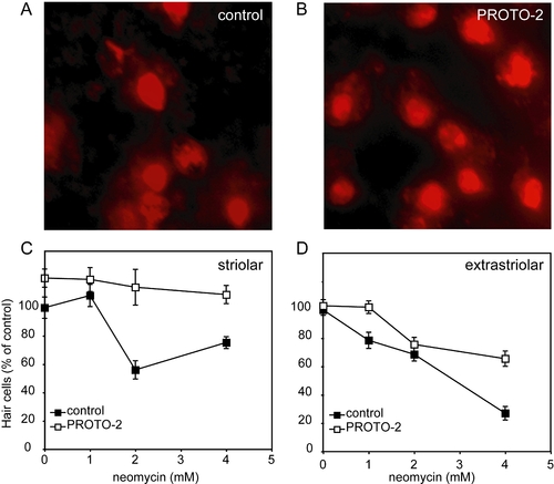
Protective compounds reduce neomycin toxicity in adult mouse utricle cultures.
(A,B) Extrastriolar utricular hair cells stained with antibodies against calmodulin and calbindin after 4 mM neomycin exposure. An increased number of hair cells remain after PROTO-2 pretreatment (B) compared to control (A). (C,D) Neomycin dose-response curve showing effect of 10 μM PROTO-2 pretreatment on striolar (C) and extrastriolar (D) utricular hair cells. Counts were made at high magnification in areas of 900 μM2, converted to density, and averaged over the three sampled areas of each region for each utricle. Ten utricles were analyzed for each treatment group. Data were normalized relative to mock-treated controls (no PROTO drug, no neomycin).

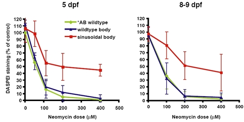
Comparison of the dose response curve of 5 dpf versus 8–9 dpf sentinel mutants. Hair cell staining by DASPEI was assessed after neomycin exposure among progeny of sentinel heterozygous parents with wildtype body shape (blue) or sinusoidal body shape (red). The dose response curves of wildtype *AB control fish are shown (green). (A) Dose-response at 5 dpf. (B) Dose-response at 8–9 dpf. Error bars are ±1 S.D.

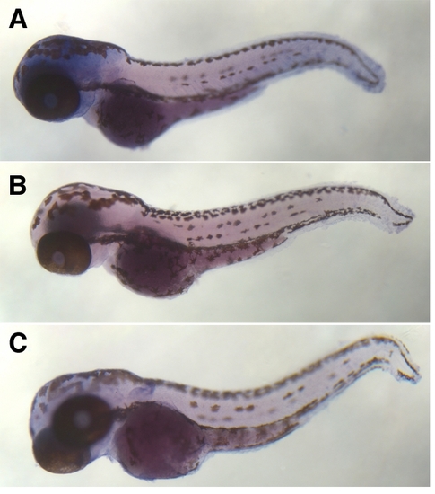
In situ hybridization of biotinylated probes to the sentinel locus reveals ubiquitous expression. (A) Wildtype *AB larvae 64 hpf, antisense probe. (B) *AB larvae 64 hpf, sense probe. (C) sentinel mutants 64 hpf, antisense probe.

EXPRESSION / LABELING:
Gene:
Fish:
Anatomical Term:
Stage: Pec-fin

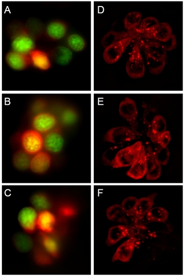
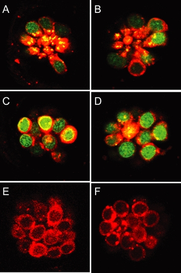
Protective compounds do not affect transduction-dependent dye or aminoglycoside uptake. (A-C) Rapid entry (45 sec) of 3 μM FM 1–43 (red) into untreated (A), PROTO-1 (B), or PROTO-2 (C) treated hair cells. Nuclei are labeled with Yo-Pro-1 (green). (D-F) Exposure to gentamicin-conjugated Texas Red results in rapid labeling of untreated (D), 10 μM PROTO-1 (E), or PROTO-2 (F) pretreated hair cells.

Unillustrated author statements

PHENOTYPE:
Fish:
Observed In:
Stage: Days 7-13
Acknowledgments
This image is the copyrighted work of the attributed author or publisher, and ZFIN has permission only to display this image to its users. Additional permissions should be obtained from the applicable author or publisher of the image. Full text @ PLoS Genet.