
- Title
-
Expression of protocadherin 18 in the CNS and pharyngeal arches of zebrafish embryos
- Authors
- Kubota, F., Murakami, T., Tajika, Y., and Yorifuji, H.
- Source
- Full text @ Int. J. Dev. Biol.
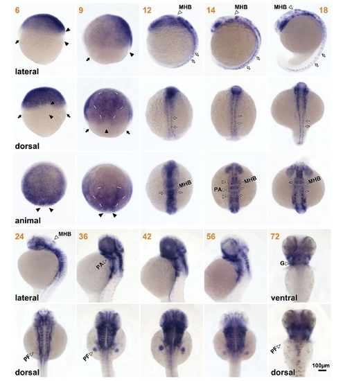
Overview of pcdh18 expression in zebrafish embryos. Panels show extended depth of field photographs (see Experimental Procedures) of embryos stained blue by in situ hybridization for pcdh18 transcripts. Orange numbers indicate hours post-fertilization. Black arrows indicate the germ ring. Black arrowheads indicate the shield. Thin white bars show the triangular area of expression in 9 hpf embryos. Open arrows indicate punctuate expression in the head and trunk. The animal pole is aligned toward the top in lateral, dorsal, and ventral views. Dorsal is to the right in lateral views and down in animal pole views. Abbreviations: G, gills; MHB, midbrain-hindbrain boundary; PA, pharyngeal arch primordia or arches; PF, pectoral fin. EXPRESSION / LABELING:
|
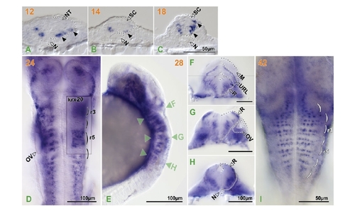
pcdh18 expression in the hindbrain and spinal cord. Orange numbers indicate hours post-fertilization. Black dotted lines outline the structures indicated. Black arrowheads in transverse sections of the trunk of 12–18 hpf embryos (A-C) (dorsal to top) indicate pcdh18-expressing cells in the lateral neural tube and spinal cord. (D) Dorsal view of a 24 hpf embryo stained for pchd18 and krx-20 with single-color double in situ hybridization. Parentheses indicate rhombomeres. Inset shows a focal plane of an area of krx-20 expression which is located ventral to that of pcdh18 expression. Green arrowheads in the lateral view of a 28-hpf embryo (E) indicate focal planes in hand-cut sections (F-H)(dorsal to top) made from the very same embryo. (F) A composite picture of two closely adjacent focal planes. (I) A dorsal view of the hindbrain (rostral to the top) of a 42 hpf embryo with patterns of transverse dotted lines. Abbreviations: M, midbrain; N, notochord; NT, neural tube; OV, otic vesicle; R, rhombencephalon; r3 & 5, rhombomere 3 & 5; SC, spinal cord; URL, upper rhombic lip. EXPRESSION / LABELING:
|
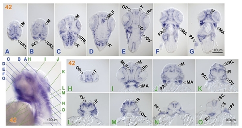
Expression in the CNS and pharyngeal arches in sections. Orange numbers indicate hours post-fertilization. Colored lines A–O in the lateral view of a 42-hpf embryo (lower left) indicate planes of serial sections (A-G) (horizontal sections; rostral to the top) and (H-O) (transverse sections; dorsal to the top). Abbreviations: 4V, forth ventricle; C, chin; D, diencephalon; E, endoderm; M, midbrain; MLF, medial longitudinal fascicle; OV, otic vesicle; OP, olfactory pit; PA, pharyngeal arches; PF, pectoral fin; R, rhombencephalon; Rn, retina; S, somite; T, telencephalon; URL, upper rhombic lip. EXPRESSION / LABELING:
|
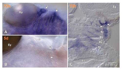
pcdh18 expression in the branchial arches. Orange numbers indicate embryonic or larval age post-fertilization. (A,B) Ventral views of embryos at 72 hpf and 5 dpf, respectively, with the yolk removed. Rostral to the left, left to the top. Dotted lines outline the jaw and branchial arches. Closed arrowheads identify the arches. (A) The 5th arch where the pharyngeal tooth would attach has diffuse but no tooth-specific pcdh18 staining at 72 hpf. (B) A pharyngeal tooth (closed arrow) appeared by 5 dpf when pcdh18 expression had diminished from the embryo. (C) A coronary cryostat section of a stained 72 hpf embryo cut at the level of the arches (the 4th arch is somewhat off the level of the particular section). The cells expressing pcdh18 (open arrowheads) surrounds branchial cartilages (open arrows). Abbreviations: 1-5, 1st to 5th branchial arches; Ey, eyes; Es, esophagial lumen, J, jaw arches. EXPRESSION / LABELING:
|