
- Title
-
Changes of gamma-tubulin expression and distribution in the zebrafish (Danio rerio) ovary, oocyte and embryo
- Authors
- Liu, J., and Lessman, C.A.
- Source
- Full text @ Gene Expr. Patterns
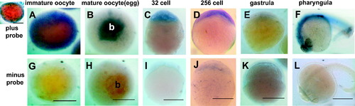
In situ hybridization (probed with FITC-oligo γ-tubulin probe) of different stages of zebrafish oocytes (A and G are fully grown, immature oocytes; B and H are mature oocytes or eggs) and embryos (C and I are 32 cell; D and J are 256 cell; E and K are ∼30% epiboly gastrula; F and L are pharyngula). The top panels are “with probe” treatments (A–F) and the lower panels are corresponding “without probe” treatments (G–L). The primordial blastodisc (b) that forms at the animal pole of the egg during oocyte meiotic maturation labels intensely with the probe (B). Inset: immature oocyte probed after hemisection showing cortical label. Anti-FITC second antibody conjugated to alkaline phosphatase and the substrate DAB were used to develop color. The specimens were dehydrated in 100% MeOH and placed in clearing media benzyl benzoate:benzyl alcohol (2:1) prior to mounting on slides. Scale bar equals 250 μm. |
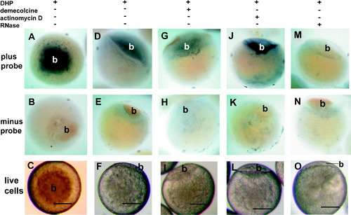
γ-Tubulin in situ hybridization of 17 α 20 β dihydroxyprogesterone (DHP 1 μg/ml) matured oocytes. γ-Tubulin message was found predominately in the animal pole primordial blastodisc (b). (A, D, G, J and M) incubated with probe; (B, E, H, K, and N) incubated without probe. (M and N) preincubated with RNase prior to hybridization. (G and H) preincubated with demecolcine (1 μg/ml) and DHP prior to fixation and hybridization. J and K preincubated with actinomycin D (1 μg/ml) and DHP prior to fixation and hybridization. (C, F, I, L, and O) are live cells showing primordial blastodisc (b) formation at the animal pole after DHP incubation. (A, B and C) are animal pole views, while all others are side views. Scale bar equals 200 μm. |
Reprinted from Gene expression patterns : GEP, 8(4), Liu, J., and Lessman, C.A., Changes of gamma-tubulin expression and distribution in the zebrafish (Danio rerio) ovary, oocyte and embryo, 237-247, Copyright (2008) with permission from Elsevier. Full text @ Gene Expr. Patterns