- Title
-
Transcription factor onecut3 regulates intrahepatic biliary development in zebrafish
- Authors
- Matthews, R.P., Lorent, K., and Pack, M.
- Source
- Full text @ Dev. Dyn.
|
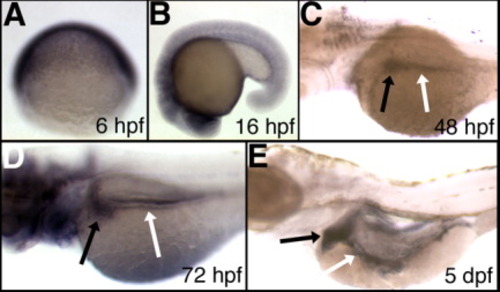
Developmental expression of zebrafish oc3. Whole-mount RNA in situ hybridizations of zebrafish embryos and larvae using an oc3 antisense probe. A: Lateral view of 6 hpf (shield stage). B-E: Left lateral views of (B) 16-hpf (18 somites), (C) 48-hpf, (D) 72-hpf, (E) 5-dpf embryos and larvae. There is low-level diffuse expression of onecut3 at 6 hpf (shield stage) (A) and 16 hpf (18 somites) (B). By 48 hpf (C), there is expression in the liver (black arrow) and intestine (white arrow), which persists at 72 hpf (D) and 5 dpf (E). |
|
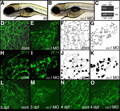
Antisense morpholino-mediated knockdown of oc3 perturbs bile duct development. A,B: Left lateral view of live 5-dpf (A) control and (B) onecut3 morpholino-injected larvae. Note the similarity in liver size and morphology of these larvae (black outline and arrow). C: Semiquantitative PCR of oc3 and tbp (TATA-box binding protein gene) using RNA from control 24-hpf larvae (cont) and from 24-hpf larvae injected with the splice-blocking oc3 morpholino, IE2 MO. Morpholino knockdown significantly reduces oc3 expression. D-G, L-O: Confocal projections of cytokeratin immunostained 5-dpf (D, E), 3-dpf (L, M), or 4-dpf (N, O) whole-mount livers injected with vehicle control (D, L, N), or oc3 morpholino antisense oligonucleotide (E, M, O). The similarity of all 3 oc3 morpholino-injected bile duct staining patterns suggests that the progression of biliary development was inhibited by the knockdown. Non-biliary cytokeratin staining in oc3 morphant livers is similar to the keratin staining seen in 3-dpf wild-type larvae (L). F, G: Schematic representations of the confocal projections in D and E. Ducts are depicted as thin lines while terminal ductules are depicted with a larger caliber in this schematic. These schematics do not account for depth of the confocal projection; thus, all ducts are rendered with equal intensity. Confocal projections of 2F11 immunostained 5-dpf whole-mount livers from control (H) and Oc3 morpholino-injected (I) larvae. There are fewer ducts and biliary cell morphology is abnormal in this morpholino-injected larva. J, K: Schematic representation of the confocal projections in H and I. Ducts are depicted as thin lines while cell bodies are the larger black structures. As with F and G, the schematics do not account for the depth of the confocal projection. Comparable ductal defects and cell morphology defects were observed in 3 other morpholino-injected larvae analyzed (not shown). PHENOTYPE:
|
|
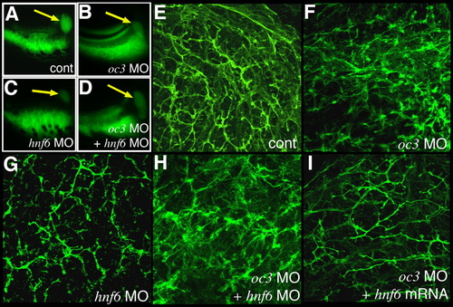
Antisense mediated knockdown of oc3 and hnf6 has a similar effect on bile duct function and development as knockdown of oc3 alone. A-D: PED6 uptake in 5-dpf larvae injected at the one-cell stage with vehicle (A), oc3 morpholino antisense oligonucleotide (B), hnf6 morpholino (C), or oc3 and hnf6 morpholinos together (D). Note that biliary secretion, measured as gallbladder fluorescence, is comparably reduced in Oc3-deficient (B), Hnf6-deficient (C), and Oc3/Hnf6-deficient larvae (D) relative to control (A). E-I: Confocal projections of cytokeratin immunostained 5-dpf whole-mount livers injected with vehicle control (E), oc3 morpholino antisense oligonucleotide (F), hnf6 morpholino (G), oc3 and hnf6 morpholinos together (H), or oc3 morpholino and hnf6 mRNA (I). The biliary phenotype of Oc3-deficient larva (F) is much more pronounced than that of the Hnf6-deficient larva (G). Rescue of the Oc3-deficient biliary phenotype by hnf6 mRNA injection is evident when I is compared to F. Note the lack of ectopic cytokeratin and the presence of intact ductular structures in I vs. F. |
|
Comparison of liver size and morphology of Oc3 morpholino-injected larvae. (A-B) Left lateral views of in situ hybridizations of 5 dpf control (A) and Oc3 morpholino-injected (B) larvae using transferrin (tf) as riboprobe. Similar results were obtained using a ceruloplasmin riboprobe (not shown). Livers are denoted by black arrows. Note the similarity of size between controls and morpholino-injected larvae. (C-D). Cross-sections of 5 dpf liver from (C) control and (D) Oc3 morpholino-injected larvae stained with hematoxylin and eosin. Tissues were fixed in paraformaldehyde, embedded in paraffin, and stained in accordance with standard techniques. Note the similarity in appearance. (E-F) Electron micrographs of control (E) and Oc3 morpholino-injected 5 dpf larvae (F). Note the overall similarity in appearance of hepatocytes, which represent almost the entirety of the depicted fields. Other structures identified include glycogen (g), liver sinusoid (s), and bile duct (bd). Scale bars denote 10 μm. EXPRESSION / LABELING:
|


