- Title
-
The serotonergic phenotype is acquired by converging genetic mechanisms within the zebrafish central nervous system
- Authors
- Lillesaar, C., Tannhauser, B., Stigloher, C., Kremmer, E., and Bally-Cuif, L.
- Source
- Full text @ Dev. Dyn.
|
A-Y: Expression of pet1 (A-L) and tph2 (M-Y) revealed by in situ hybridization on whole-mount embryos/larvae (A-J and M-V) or brains (K-L and X-Y). Insets show higher magnifications of the boxed areas. The earliest pet1-positive cells are likely adrenal gland (Aa′ and Bb″) and blood (Aa″) precursors. B: The first pet1-expressing cells in the rhombencephalon are detected at 25 hours postfertilization (hpf). O: Expression of tph2 in this location is not detected until a few hours later. Afterward, pet1 and tph2 expression highlights two parallel stripes of precursors lining the hindbrain floor plate. E,G,I,S,U: These are organized in an anterior (arrowhead) and posterior (arrow) cluster separated by a gap (white arrow). H: An additional column of pet1-positive cells runs along this domain in a slightly more dorsal location (stars in h′). Starting from 60 hpf tph2 expression can also be seen in an additional cluster of cells corresponding to the pretectal complex (Kaslin and Panula,[2001]). In addition, tph2 was detected in the epiphysis at all stages examined. e, epiphysis; ptc, pretectal and thalamic complex. EXPRESSION / LABELING:
|
|
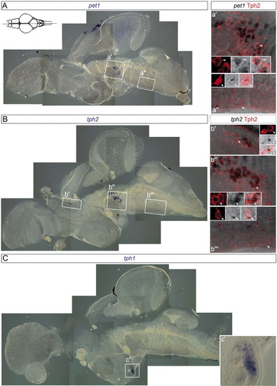
Comparison of the localization of pet1, tph2, and tph1 transcripts (in situ hybridization, blue/black) and of Tph2 protein (immunohistochemistry, red) on adult brain sagittal sections. A: In situ staining for pet1 in anterior (a′) and posterior (a″) raphe nuclei. a′ and a″ (optical projections) show higher magnification of boxed areas in A together with Tph2 immunostaining in red. Note the double-labeled cells, some of which (arrowheads) are further magnified in the small insets (optical sections). B: In situ staining for tph2 in anterior (b″) and posterior (b″′) raphe nuclei and in the pretectal complex (b′). b′, b″, and b″′ (optical projections) show higher magnification of boxed areas in B together with Tph2 immunostaining. Double labeling was observed in all three regions for all cells, some of which (arrowheads) are further magnified in the small insets (optical sections). C: In situ staining for tph1 in the hypothalamus. c′ shows higher magnification of boxed area in C. Schematic picture was modified from Wullimann et al. ([1996]). EXPRESSION / LABELING:
|
|
Compared localization of pet1 and tph2 transcripts (in situ hybridization, black/blue) and of Tph2 protein (immunohistochemistry, red) on adult brain coronal sections. Arrowheads indicate examples of double-labeled cells. A,B:pet1 transcripts and Tph2 protein shown in optical projections of sections through the dorsal and the medial raphe, respectively, at the level indicated in the schematic pictures. Color brightfield pictures were included (right panels) to clarify the distinction between cross-cut fiber bundles (that appear dark on black-white images, but are negative for pet1 transcripts) and blue in situ staining. The dorsal raphe (corresponding to cluster B6-B7) is located more dorsally and laterally than the medial raphe (B8-B9; Kaslin and Panula,[2001]). C-E: The location of tph2 transcripts and Tph2 protein in the pretectal complex (level of section indicated in schematic picture) is illustrated (C) in addition to the dorsal (D) and median (E) raphe in optical projections. Schematic picture was modified from Wulliman et al. ([1996]). EXPRESSION / LABELING:
|
|
Location of the gap separating the anterior and posterior pet1-positive raphe precursors in relation to green fluorescent protein (GFP) -positive cell clusters in the isl1:gfp transgenic line. Photomicrographs are confocal optical projections of dorsal views at hindbrain levels, anterior left. A: Mauthner neurons (arrows), located in rhombomere (r) 4, were labeled by retrograde tracing in 6 days postfertilization (dpf) isl1:gfp transgenic larvae using rhodamine dextran. B,C: Thereby, the spatial distribution of GFP-expressing cells (B, arrowheads) in relation to rhombomeres was determined (C, overlay of A and B). C: Note that the Mauthner neurons overlap with a cluster of GFP-positive cells (*) just posterior to the trigeminal nuclei (Va and Vp, arrows). D: The anterior and posterior clusters of serotonergic precursors were identified using an antibody against tryptophan hydroxylase (Tph) 2. The inset shows a higher magnification of the boxed area, and white arrowheads in the inset indicate the gap between the anterior and posterior Tph-positive clusters (optical section). E,F: Note, in F (overlay of D and E) that this gap overlaps with Vp, in r3 (black arrowheads), rather than with Mauthner neurons, in r4. Thus, the anterior Tph cluster spans r1-2, and the posterior cluster spans r4 and beyond. |
|
Origin of rhombomere (r) 1-2 serotonergic precursors. A: Strategy for tracing the origin of r1-2 serotonergic precursors in her5PAC:egfp transgenic embryos injected with caged-fluorescein at one-cell stage (dorsal views, anterior left). An ultraviolet-light beam was focused along the midline (i) within or (ii) posterior to the green fluorescent protein (GFP) -expressing area at 90% epiboly/tail bud (left drawing; green GFP-positive midbrain-hindbrain boundary [MHB] progenitor pool). Photomicrographs: control embryos fixed directly after uncaging and processed for GFP (green) and fluorescein (red) immunostaining. Schematic pictures: two possible outcomes at 36 hours postfertilization (hpf). Top: uncaging within the GFP-positive area and pet1-positive cells of r1-2 fluorescein-labeled (red); origin within the MHB pool; Bottom: uncaging posterior to the GFP-positive area and pet1-positive cells of r1-2 fluorescein-labeled; origin posterior to the MHB pool. B: A 16-μm optical projection of a coronal cryosection through r1-2 of embryos uncaged within the GFP-expressing domain. Insets: High magnifications of 1-μm optical section of double-labeled cell indicated with an arrow. Arrowheads: pet1-positive cells on each side of the floor plate within r1-2. Note anti-fluorescein labeling of these pet1-positive cells. C: Same analysis in an embryo uncaged posterior to the GFP-expressing domain. mes, mesencephalon. Schematic picture modified from Mueller and Wullimann ([2005]). |


