
- Title
-
A dual role for the zebrafish unplugged gene in motor axon pathfinding and pharyngeal development
- Authors
- Zhang, J., Malayaman, S., Davis, C., and Granato, M.
- Source
- Full text @ Dev. Biol.
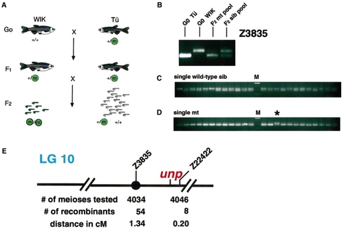
Molecular-genetic mapping of the unplugged locus to LG 10. (A) Schematic drawing showing the three-generation mapping cross. In the G0 generation, a heterozygous unplugged fish in the Tü background was crossed to a polymorphic WIK fish. F1 heterozygous fish were crossed to each other, F2 homozygous mutants and their siblings were collected, and their DNA was extracted for chromosomal mapping. (B) PCR amplification using marker Z3835 and DNA isolated from the G0 Tü fish, the G0 WIK fish, pooled F2 mutants, and pooled F2 siblings. The Tü -specific PCR fragment is shorter than the WIK-specific fragment. Amplification with Z3835 in F2 sibling pool DNA results in a Tü -specific and a WIK-specific fragment, while in DNA from pooled mutant only the Tü -specific fragment is present (higher mobility), suggesting linkage between Z3835 and the unplugged mutation. (C) PCR amplification using marker Z3835 and DNA from individual F2 sibling embryos. Embryos in which only a WIK-specific fragment is present (lower mobility) are of +/+ genotype, while embryos with a Tü -specific and a WIK-specific PCR fragment are heterozygotes (+/- genotype). (D) PCR amplification using marker Z3835 and DNA from individual F2 unplugged mutant embryos. In most mutant embryos, only the Tü -specific fragment is present. In recombinant embryos, a Tü -specific and a WIK-specific fragment are present (*). In these embryos, the unplugged Tü chromosome recombined with the WIK chromosome between Z3835 and the unplugged locus. (E) Schematic representation of LG 10 around the unplugged locus. The centromere is indicated by a black circle. |
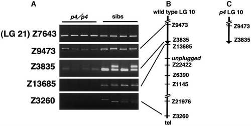
The unplugged locus is absent in p4 mutants. (A) PCR amplification of SSLP markers from DNA of four p4/p4 mutant embryos and four sibling embryos. In sibling embryos (sibs), all tested markers are present. In the four p4/p4 embryos, LG 21 marker Z7643 and LG 10 markers Z9473 and Z3835 are present, while markers Z13685 and Z3260 (telomeric) are absent. (B) Schematic representation of LG 10 around the unplugged locus. Relevant SSLP markers located between the centromere (black circle) and the telomere are indicated. (C) In p4/p4 mutant embryos, LG 10 breaks between Z3835 and Z13685, deleting the entire chromosomal arm, including the unplugged locus. |
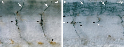
unplugged mutant embryos display specific motor axon defects at the choice point. Lateral views of 27-hpf wild-type (A) and unplugged mutant (B) embryos stained with znp-1 antibody. The common path extends between the lower end of the spinal cord (black line) and the horizontal myoseptum (white line). (A) In wild-type embryos, CaP axons (black arrow) and MiP axons (white arrow) have completed migration on the common path and extend on their cell-type specific trajectories. (B) In unplugged mutant embryos, MiP trajectories are unaffected (white arrows). In contrast, presumptive RoP and CaP motor neurons display two classes of axonal phenotypes at the choice point (black arrowhead). Mutant motor axons stalled at the choice point instead of extending further into the ventral myotome (stall phenotype, white arrowhead), or they extended into the ventral myotome but formed ectopic branches at the choice point (branching phenotype, black arrow). |
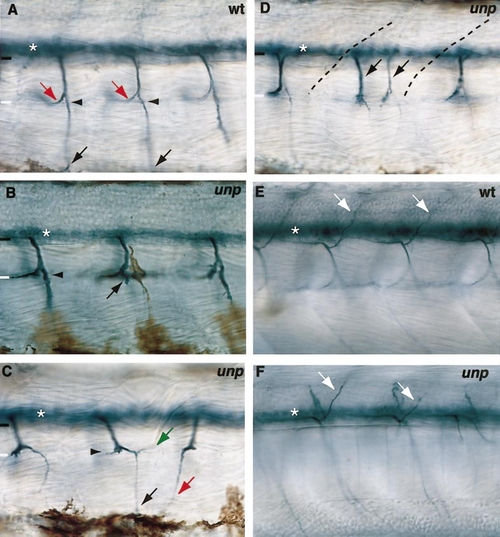
unplugged mutant embryos exhibit multiple secondary motor axon defects. Lateral views of wild-type (A, E) and unplugged mutant (B–D, F) embryos stained with zn-5 antibody at 45 (A–D) or 60 hpf (E, F). The zn-5 antibody recognizes cell bodies (white asterisk in A–F) and axons of secondary but not primary motor neurons. The common path is found between the lower end of the spinal cord (black line) and the horizontal myoseptum (white line). (A) In wild-type embryos, a ventral nerve extends into the ventral myotome (black arrow), a medial nerve extends rostrally along the horizontal myoseptum (red arrow), and a dorsal nerve projects into the dorsal myotome (white arrow in E). Note that the medial nerve defasciculates from the ventral root before reaching the choice point (black arrowhead), and then turns rostrally and projects along the horizontal myoseptum (red arrows in A). Ventral nerves stalled at the choice point (black arrow in B) or projected into the ventral myotome, but form an aberrant branch (green arrow) at the choice point (C). In a significant fraction of hemisegments the medial nerve invaded the ventral myotome (red arrow in C). (D) In 3–6% of unplugged somitic hemisegments, secondary motor axons exit the spinal cord as two independent trajectories (black arrows; somite boundaries are indicated by dashed lines). (E) At ∼60 hpf, zn-5 stains the dorsal nerves of secondary motor axons (white arrows). (F) In unplugged mutant embryos, dorsal nerves appear indistinguishable from those in wild-type embryos. |
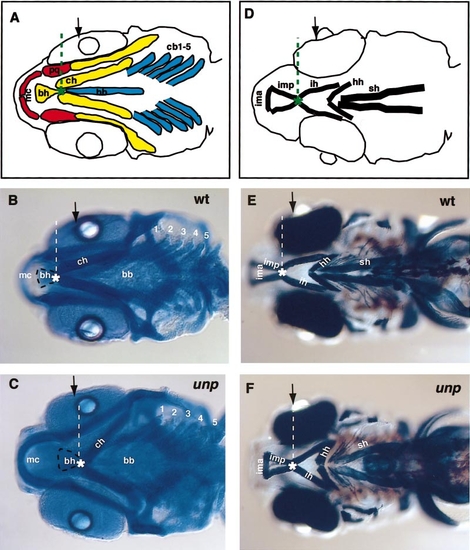
Pharyngeal arch defects in unplugged larvae. (A) Schematic drawing of pharyngeal arch cartilages. The components of the mandibular arch are labeled in red, the hyoid arch in yellow and the five branchial arches in blue. Note that not all cartilage components are indicated, and that only the distal portions of the branchial arches are outlined. The contact point (green asterisk) between the ceratohyals (ch) and the basihyal (bh) is located anterior to the lens (black arrow). (B, C) Five-dpf wild-type and unplugged mutant larvae stained with Alcian blue. (B) In wild-type larvae, the contact point between the basihyal (outlined by a black dashed line) and the ceratohyals (white asterisk) is located anterior to the lens (black arrow). Note that Meckel’s cartilages are situated further dorsally than the six posterior arches, and therefore appear slightly out of focus. (C) In unplugged mutants, Meckel’s cartilages (mc) are displaced ventrally to the same dorso-ventral position as the posterior arches, and now appear in the same focal plane. Also, the basihyal and the anterior end of the ceratohyals are shifted more posteriorly. As a result, the basihyal now contacts the ceratohyals at the level of the lens (dashed line). (D) Schematic representation of pharyngeal muscles. Note that the contact point (green asterisk) between the interhyal (ih) muscle and the intermandibularis posterior (imp) muscle is located anterior to the lens (black arrow). (E, F) Five-dfp wild-type and unplugged mutant larvae stained with the MF-20 antibody for pharyngeal muscles. Note that in wild-type larvae, the interhyal (ih) muscle contacts the intermandibularis posterior (imp) muscle at a level anterior the lens (black arrow). In unplugged mutant larvae, the interhyal (ih) muscle contacts the intermandibularis posterior (imp) muscle at a more posterior position, at level of the lens (black arrow in F). Abbreviations: bb, basibranchials; bh, basihyal; cb1-5, ceratobranchial 1-5; ch, ceratohyal; hh, hyohyal; ih, interhyal; ima, intermandibularis anterior; imp, intermandibularis posterior; mc, Meckel’s cartilage; pq, palatoquadrate; sh, sternohyoideus. |
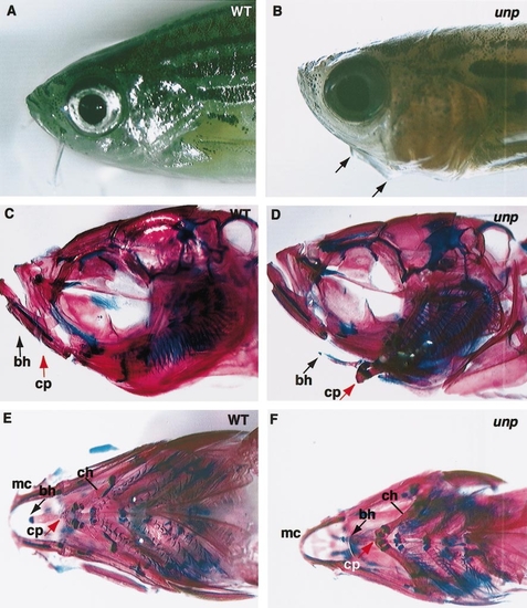
Pharyngeal arch defects in unplugged mutant adult fish. Lateral views of 17-week-old wild-type (A) and unplugged mutant (B) fish. In unplugged mutant fish (B), a ventral protrusion is visible from the lower jaw (black arrows in B). Skeletal preparations of the same wild-type (C, E) and unplugged mutant (D, F) adult fish. Lateral (C, D) and ventral (E, F) views of wild-type (C, E) and unplugged fish stained for chondrocytes (blue) and calcified bones (red). (C) In wild-type fish, the basihyal (bh; black arrow) and its connection point (cp; red arrow) with the ceratohyals (ch) are not distinguishable in a lateral view, and only their approximate locations are indicated. (D) In unplugged mutant fish, the basihyal (black arrow) and its connection point with the ceratohyals (red arrow) are located more ventrally and posteriorly. (E) Ventral view of the same wild-type preparation shown in C. (F) Ventral view of the same unplugged preparation shown in D. Note the increased distance between Meckel’s cartilages (mc) and the anterior tip of the basihyal (bh, black arrow), compared to wild-type fish (E). cp, connection point between the ceratohyals and the basihyal; bh, basihyal; ch, ceratohyal; mc, Meckel’s cartilages. |
Reprinted from Developmental Biology, 240(2), Zhang, J., Malayaman, S., Davis, C., and Granato, M., A dual role for the zebrafish unplugged gene in motor axon pathfinding and pharyngeal development, 560-573, Copyright (2001) with permission from Elsevier. Full text @ Dev. Biol.