- Title
-
The expression of a zebrafish gene homologous to Drosophila snail suggests a conserved function in invertebrate and vertebrate gastrulation
- Authors
- Hammerschmidt, M. and Nüsslein-Volhard, C.
- Source
- Full text @ Development
|
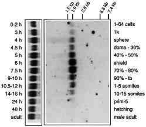
Temporal expression of sna-1 mRNA. 10 mg of total RNA of the indicated stages were blotted. Staging was done according to Westerfield (1993). A single transcript of about 1.9 kb was detected upon probing with the 933 bp BamHI-HindIII fragment of pBS-Sn3. The left panel shows methylene blue staining of ribosomal RNA of the same blot. |
|
(A) Specificity of anti-Sna-1 antibodies. Coomassie-bluestained SDS-PAGE gel (lanes A, B) and Western blot (lanes C-F) showing total protein from induced (lanes A,D) and uninduced bacteria (lane C) carrying expression vector pQE12-sn, and Ni- NBT purified protein from pQE12-sn containing (lanes B,E) and pQE16-snD114 containing bacteria (lane F). The band with an apparent Mr of 39x103 was used to raise the antiserum. (B) Temporal expression pattern of Sna-1 protein. Western blot showing Ni-NBT-purified protein of pQE12-sn carrying bacteria (lane H) and total protein equivalent to 2-3 zebrafish embryos of following stages: 1-64 cells (lane A), sphere (lane B), shield (lane D), tailbud (lane E), 1 day (lane F) and 6 days (lane G). The zebrafish Sna-1 band was identified by comparison of the protein pattern of sphere-stage embryos, which were either untreated (lane B) or injected with pBS-sn3 encoded sna-1 sense RNA in the 1-cell stage (lane C). The injection leads to the exclusive increase of one band of an apparent Mr of 39x103 which comigrates with the pQE12-sn expressed recombinant protein. |
|
Expression of sna-1 in wild-type embryos before and during gastrulation, investigated by whole-mount in situ hybridisation (A,C,D,E,F,G,H) and anti-Sna-1 antibody staining (B). (A) Sphere stage, lateral view; (B) sphere stage, animal view, counterstained with DAPI; (C) dome stage, animal view, dorsal right; (D) 30% epiboly, lateral view, dorsal right; (E) germ ring stage, animal view, dorsal right; (F) germ ring stage; lateral view; (G) 60% epiboly, dorsal view; (H) 80% epiboly, dorsal view. Abbreviations: epiblast (e), hypoblast (h). For details see text. EXPRESSION / LABELING:
|
|
Expression of sna-1 in wild-type, spt and ntl mutant postgastrula embryos, investigated by whole-mount in situ hybridisation (A,B,C,D,E,G,H,J,L,M) and anti-Sna-1 antibody staining (E, small photo, F,K). (A,B) 95% epiboly, dorsal view: (A) wild-type, (B) ntl. (C,D) 4-somite stage, optical cross section at level of presomitic mesoderm: (C) wild-type, (D) ntl. (E, small photo) 5-somite stage, dorsal view on somites. (F) 15-somite stage, spread wild-type embryo, dorsal view on posterior part of the embryo up to somite 12. (E, big photo, G,H) 8-somite stage, dorsal view, spread embryo: (E) wild type; (G) ntl ; (H) spt. (K) 15-somite stage, wild type, lateral view on somites 10-13. (J,L,M) 22 hour embryos, lateral view of the tail and the posterior part of the trunk; borders of embryo outlined by dots: (J) wildtype; (L) ntl ; (M) spt. Abbreviations: muscle pioneer precursor (mpp), notochord (n), somite (s), tailbud (tb). For details see text. EXPRESSION / LABELING:
|
|
Expression of sna-1 in the head region and the pectoral fin buds investigated by whole-mount in situ hybridisation. (A) 1 day, lateral view; sna-1 is expressed in the tip of the tail, the posterior somites and in ventral lateral regions of the head sparing the trigeminal ganglion and the otic placode. (B,C) 2 days, lateral view; sna-1 is predominantly expressed in lateral regions of mandibular and hyoid (B), and in medial parts of all arches including the 5 gill arches (C). (D) 2.5 days, dorsal view; sna-1 expression in the two presumptive muscle bundles of the pectoral fin bud. Abbreviations: trigeminal ganglion (tg), otic placode (op), somites (s), mandibular (md), hyoid (hy), gill arches (ga), pectoral fin bud (pfb). |
|
Relationship of sna-1 and ntl expression in wild-type (C-H) and ntl mutant (A,B) embryos investigated by sna-1 in situ (blue) and anti-Ntl antibody (brown) double stainings. All embryos were spread. (A,D) Germ ring stage, dorsal view: (A) ntl, (D) wild-type. (C) Section through germ ring of germ ring-stage embryo, lateral position. The border of the embryo and the separation of epiblast and hypoblast are outlined by dots. (B,E) 60% epiboly, dorsal view: (B) ntl, (E) wild type. (F) 80% epiboly, dorsal view.(G) 95% epiboly, dorsal view. (H) 4-somite stage, dorsal view. For details see text. Arrows in E and F indicate the ?forerunning? cells, a group of sna-1- negative and ntl-positive cells located on the yolk posterior to the germ ring at the dorsal midline of the embryo. EXPRESSION / LABELING:
|
|
sna-1 expression after ectopic expression of ntl in intact embryos (A,B; stained for Ntl protein and sna-1 mRNA) and after activin A treatment of animal caps (C,D; stained for Sna-1 protein). (A) Germ ring-stage embryo. Ntl protein is ectopically expressed in animal regions of the embryo. sna-1 expression is restricted to the germ ring, its endogenous expression domain. (B) Sphere stage. Ntl protein is ectopically expressed and overexpressed in the right part of the embryo (separated by dashes). sna-1 is more strongly expressed at the margin of the right part of the embryo. (C) Expression of Sna-1 protein upon incubation of the animal cap for 5 hours in medium containing activin A. (D) No Sna-1 expression upon incubation in control medium. The animal caps are slightly spread. |


