
- Title
-
Germ cells are required to maintain a stable sexual phenotype in adult zebrafish
- Authors
- Dranow, D.B., Tucker, R.P., and Draper, B.W.
- Source
- Full text @ Dev. Biol.

Phenotypic sex reversal of nanos3 -/- 1° females correlates with loss of oocytes. (A-A′′) Representative 2.5 month old (mo) nanos3 +/- female control. (B-B′′) nanos3 -/- 1° female with large, egg-filled abdomen and light yellow fin pigmentation characteristic of control females. (C-C′′) Representative 5 mo nanos3 -/- 1° female. (D-D′′) Representative 5 mo nanos3 +/- male control. (E-E′′) Representative 5 mo nanos3 +/- female. At 5 mo, nanos3 -/- 1° females (C) have slim bodies and darker yellow fin pigmentation characteristic of control males (D). (A-E) Low magnification pictures showing anterior body. (A′-E′) Higher magnification picture showing anal fin. (A′′-E′′) magnified view of regions boxed in A′-E′. See also Fig. S1. |

Germ cell ablation in adult zebrafish induces sex reversal. (A-A′′) Representative DMSO-treated Tg(ziwi:CFP-NTR) female control and pre-treatment Tg(ziwi:CFP-NTR) 1° female (B-B′′) have large, egg-filled abdomens and light yellow fin pigmentation characteristic of wild-type females. (C-C′′) The same female depicted in B two months post Mtz treatment has a slimmer body and darker yellow fin pigmentation characteristic of a control male (D-D′′). (A-D) Low magnification pictures showing anterior body. (A′-D′) higher magnification picture showing anal fin. (A′′-D′′) magnified view of regions boxed in A′-D′. See also Fig. S2. |
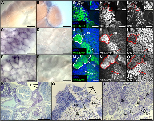
Oocyte ablation in adult zebrafish induces complete ovary-to-testis reprogramming. (A-F) in situ hybridization of wild-type (WT) and sex-reverted gonads for genes differentially expressed in the male or female somatic gonad. (A) amh is not detected in WT ovaries. (B) cyp19a1a is expressed in granulosa cells. (C) amh is expressed in Sertoli cells of the testis while cyp19a1a (D) is not detected. (E) amh but not cyp19a1a (F) is expressed in testes of sex-reverted Tg(ziwi:CFP-NTR) females. (G-O) expression of CFP-NTR in control ovary, control testis, and testis of sex-reverted fish. (G, J, and M) merged images: CFP-NTR in green, DNA in blue. (H, K, and N) CFP-NTR only. (I, L, and O) DNA only. (G-I) CFP-NTR is expressed in all germ cells of the control ovary. IAz, IAp, IAd, stage IA oocytes -- zygotene, pachytene, diplotene stages of meiotic prophase, IB, stage IB oocytes. Premeiotic germ cells (boxed and inset). (J-L) CFP-NTR expression in testis tissue from sex-reverted fish. (M-O) CFP-NTR expression in control testis. Morphology of control ovary (P), control testis (Q), and testis of sex-reverted fish (R). Sg, spermatogonia; Sc, spermatocytes; Sz, spermatozoa; S, Sertoli cells; L, Leydig cells; My, peritubular myoid cells. Scale bars: 200 μM (A-F, and P), 50 μM (G-I, Q, and R), 10 μM (G-I, insets), and 20 μM (J-O). See also Fig. S3. |

5 mo nanos3 -/- 1° females lack a prominent cloaca.(A-E) Anal fins of nanos3 fish with cloaca boxed and magnified in bottom right of each panel. Dotted lines delineate the cloaca. (A) 2.5 mo nanos3+/- female. (B) 2.5 mo nanos3-/- 1° female. (C) 5 mo nanos3 -/- 1° female. (D) 5 mo nanos3+/- female. (E) 5 mo nanos3+/- male. Image contrast adjusted for clarity. |

Mtz-treated 1° females lack a prominent cloaca (A-D) Anal fins of Tg(ziwi:CFP-NTR) fish with cloaca boxed and magnified in bottom right of each panel. Dotted lines delineate the cloaca. (A) DMSO-treated Tg(ziwi:CFP-NTR) female. (B) Pre-treatment Tg(ziwi:CFP-NTR) 1° female. (C) 2 mo post-treatment Tg(ziwi:CFP-NTR) 1° female. (D) Control Tg(ziwi:CFP-NTR) male. Image contrast adjusted for clarity. |
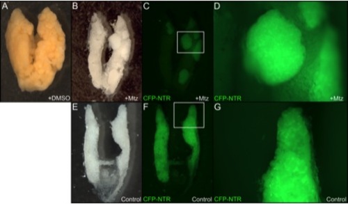
Gonads from sex-reverted Tg(ziwi:CFP-NTR) females contain testis-like tissue. (A) Ovaries of control DMSO-treated females have many large, yolky oocytes. (B) Gonads of Mtz-treated sex-reversed fish contain mostly adipose tissue with patches of opaque testis-like tissue, most similar to the testes of a control male (E). (C, D, F, G) Fluorescent micrographs of the gonad of an Mtz-treated 1° female and testes of control male with CFP pseudocolored in green. (C) Gonads from Mtz-treated females contain patches of CFP-positive cells that resemble control testis tissue (F). (D, G) Magnified views of the boxed regions in C and F, respectively. |
Reprinted from Developmental Biology, 376(1), Dranow, D.B., Tucker, R.P., and Draper, B.W., Germ cells are required to maintain a stable sexual phenotype in adult zebrafish, 43-50, Copyright (2013) with permission from Elsevier. Full text @ Dev. Biol.