
- Title
-
Progenitor expansion in apc mutants is mediated by Jak/Stat signaling
- Authors
- Lin, J., Wang, X., and Dorsky, R.I.
- Source
- Full text @ BMC Dev. Biol.

stat3 is a direct Lef1 target gene. (A) Density plot from Lef1 ChIP-seq. Sequences from total input chromatin are on top and Lef1 ChIP are below, plotted on the UCSC genome assembly. A peak of Lef1 binding is observed at the stat3 promoter (arrow). (B) Genomic sequence upstream of stat3 transcription start site, identified by Lef1 ChIP-seq. Putative Lef/Tcf binding sites are in red, and mRNA sequence is in green. (C) Direct ChIP using Lef1 antibody with primers indicated in panel (B). Mean percent of total input chromatin by qPCR from 3 independent experiments is shown. Precipitation from wild-type 36 hpf chromatin is significantly enriched over chromatin from lef1 deletion mutant embryos. ChIP from a control genomic region without putative binding sites shows no enrichment. Error bars = s.d., *p < 0.05 by student′s t-test. (D,E) stat3 expression is decreased 8 hours after ubiquitous expression of the constitutive repressor ΔTcf. Cross-sections through the hypothalamus are shown in (E). Both wild-type embryos (left) and siblings expressing hs:Δtcf (right) were heat-shocked at 28 hpf and processed for stat3 in situ hybridization at 36 hpf. |
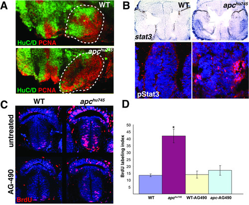
Jak/Stat signaling mediates proliferation in the apc mutant hypothalamus. (A) Proliferating cells are increased and neurogenesis is decreased in the apc mutant hypothalamus. Lateral confocal projections are shown with the neuronal marker HuC/D in green and the proliferation marker PCNA in red. Anterior is to the left and the hypothalamus is indicated by the dotted lines. (B) Both stat3 mRNA expression and pStat3 levels are increased in the apc mutant hypothalamus at 36 hpf. In situ hybridization for stat3 mRNA (top) and immunohistochemistry for pStat3 (bottom) are shown in transverse cryosections. (C) BrdU incorporation in the hypothalamus is increased in apc mutants, and restored to wild-type levels by AG-490 treatment. Embryos were labeled with BrdU for 1 hour and fixed for analysis at 36 hpf. Confocal projections from the ventral brain surface through the 36 hpf hypothalamus are shown. BrdU immunohistochemistry is red, TO-PRO-3 nuclear stain is in blue. (D) The BrdU labeling index is significantly increased in the apc mutant hypothalamus compared to wild-type siblings, and restored to wild-type levels by AG-490 treatment. Error bars = s.d., p < 0.05 by student′s t-test. |
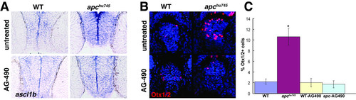
Jak/Stat signaling mediates progenitor marker expression in the apc mutant hypothalamus. (A) Expression of ascl1b, a neural progenitor marker, is qualitatively increased in the apc mutant hypothalamus, and restored to wild-type levels by AG-490 treatment. Transverse cryosections at 36 hpf are shown. (B) Expression of Otx1/2, a hypothalamic progenitor marker, is increased in the apc mutant hypothalamus, and restored to wild-type levels by AG-490 treatment. Confocal projections from the ventral brain surface through the 36 hpf hypothalamus are shown. Otx1/2 immunohistochemistry is red, TO-PRO-3 nuclear stain is in blue. (C) The percent of Otx1/2+ cells is significantly increased in the apc mutant hypothalamus compared to wild-type siblings, and restored to wild-type levels by AG-490 treatment. Error bars = s.d., p < 0.05 by student′s t-test. |
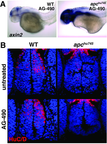
Jak/Stat signaling does generally mediate Wnt-responsive gene expression or the entire neurogenesis program in apc mutants. (A) axin2 mRNA expression, a general marker for Wnt/β-catenin target gene activation, is increased in apc mutants treated with AG-490 at 36 hpf, compared to controls. Lateral whole-mount views at 36 hpf are shown. (B) Expression of HuC/D, a marker of differentiated neurons, is decreased in the apc mutant hypothalamus, and remains decreased after AG-490 treatment. Confocal projections from the ventral brain surface through the 36 hpf hypothalamus are shown. HuC/D immunohistochemistry is red, TO-PRO-3 nuclear stain is in blue. |