
|
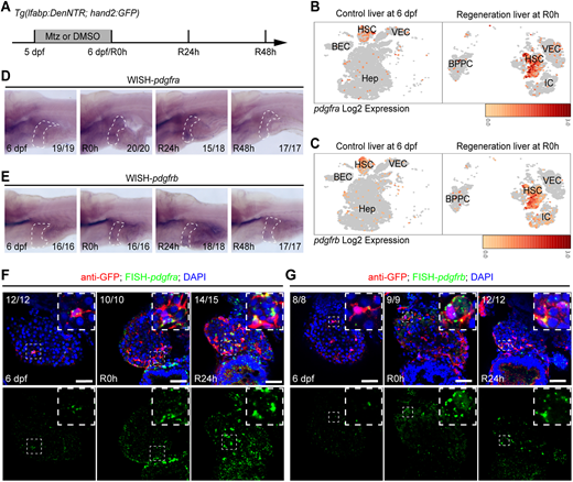
Fig. 1 PDGF receptors are upregulated in HSCs during liver regeneration. (A) Experimental scheme illustrating the regenerative phase following liver injury induced by Mtz. (B,C) UMAP showing pdgfra (B) and pdgfrb (C) expression overlaid (red) in various cell types of livers at 6 dpf and immediately after injury (at regeneration 0 h; R0 h). BEC, biliary epithelial cell; HSC, hepatic stellate cell; VEC, vascular endothelial cell; Hep, hepatocyte; BPPC, bipotential progenitor cell; IC, immune cell. (D,E) Lateral view whole-mount in situ hybridization images showing the expression of pdgfra (D) and pdgfrb (E) at 6 dpf, R0 h, R24 h and R48 h. The liver region is outlined with a dashed line. (F,G) Fluorescent in situ hybridization and antibody staining demonstrating pdgfra (F) and pdgfrb (G) expression in GFP+ HSCs at 6 dpf, R0 h and R24 h under the Tg(lfabp:DenNTR; hand2:GFP) background. Scale bars: 50 μm. Numbers indicate the proportion of larvae exhibiting the expression shown.