
Fig. 4
- ID
- ZDB-IMAGE-251103-25
- Genes
- Publication
- Sartori et al., 2025 - Zebrafish model reveals developmental and hematopoietic functions of ADAMTS13
- All Figures
- Figures for Sartori et al., 2025
|
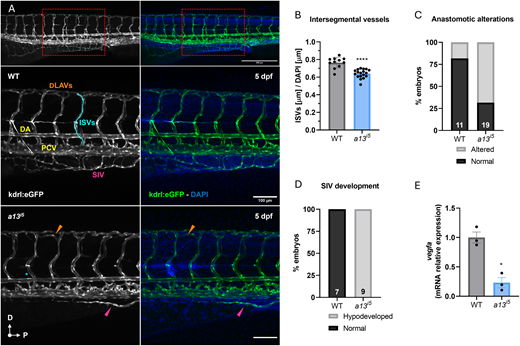
Fig. 4 ADAMTS13 deficiency impairs vascular developmental patterning. (A) Representative confocal images of the trunk vasculature of casper;kdrl:eGFP transgenic zebrafish at 5 dpf. Top panels show an overview of the tail vasculature (10×), the red rectangle highlights the area of the trunk considered for the analysis. Scale bar: 300 μm. Mid panels show a WT embryo, kdrl:eGFP in grey (left), and the merge with DAPI (right). Lower panels show an a13i5 mutant embryo, kdrl:eGFP in grey (left) and the merge with DAPI (right). Scale bar: 100 μm. (B) Quantification of ISV length (µm) normalized to DAPI in WT (grey) and a13i5 (blue) embryos. (C) Percentage of embryos with normal (black) or altered (grey) anastomotic connections in WT and a13i5 mutants. (D) Percentage of embryos with normal (black) or hypodeveloped (grey) SIV plexus in WT and a13i5 mutants. (E) Relative mRNA expression levels of vegfa in WT and a13i5 embryos, normalized to the housekeeping gene ube2a, and shown as fold versus WT. Data are shown as mean±s.d. Each dot represents a biological replicate. *P≤0.05, **** P≤0.0001 by unpaired Student's t-test. DA, dorsal aorta; DLAV, dorsal longitudinal anastomotic vessel; ISV, intersegmental vessel; PCV, posterior cardinal vein; SIV, subintestinal vein.