
Fig. 1
- ID
- ZDB-IMAGE-250917-56
- Genes
- Publication
- Tevar et al., 2025 - Zebrafish adamtsl4 knockout recapitulates key features of human ADAMTSL4-related diseases: a gene involved in extracellular matrix organization, cell junctions and development
- All Figures
- Figures for Tevar et al., 2025
|
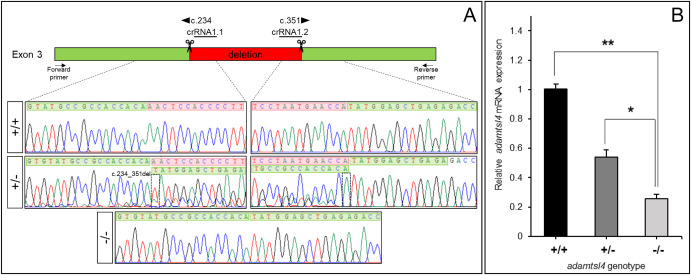
Fig. 1 Molecular characterization of the adamtsl4-KO zebrafish line generated using CRISPR/Cas9 genome editing. A. Localization of the deletion identified in adamtsl4 exon 3. Scissors: DNA cleavage sites targeted by two crRNAs (crRNA1.1 and crRNA1.2). The numbers indicate the cDNA nucleotide positions corresponding to the start and end of the deleted DNA fragment. Wild-type sequences are highlighted with a green background, while deleted sequences are marked with a red background. Arrows: position of the forward and reverse primers used for zebrafish genotyping. Rectangles in the electropherograms mark the 5′ and 3′ ends of the deletion. B. Reduced mRNA levels in adamtsl4-KO zebrafish larvae. Relative adamtsl4 mRNA levels were measured in 6 days post-fertilization (dpf) zebrafish larvae using RT-qPCR. Data represent the mean ± standard error, derived from three independent experiments performed in triplicate, using pools of 50 larvae. Statistically significant differences between KO and wild-type zebrafish are denoted by asterisks: p < 0.05 (∗), p < 0.01 (∗∗). (For interpretation of the references to colour in this figure legend, the reader is referred to the Web version of this article.)
Reprinted from Experimental Eye Research, , Tevar, A., Aroca-Aguilar, J.D., Atiénzar-Aroca, R., Ramírez, A.I., Fernández-Albarral, J.A., Escribano, J., Zebrafish adamtsl4 knockout recapitulates key features of human ADAMTSL4-related diseases: a gene involved in extracellular matrix organization, cell junctions and development, 110572, Copyright (2025) with permission from Elsevier. Full text @ Exp. Eye. Res.